- 靶点:
- APAF1
- 简介:
- >>Platinum drug resistance;>>p53 signaling pathway;>>Apoptosis;>>Apoptosis - multiple species;>>Alzheimer disease;>>Parkinson disease;>>Amyotrophic lateral sclerosis;>>Huntington disease;>>Prion disease;>>Pathways of neurodegeneration - multiple diseases;>>Legionellosis;>>Tuberculosis;>>Hepatitis C;>>Hepatitis B;>>Measles;>>Influenza A;>>Herpes simplex virus 1 infection;>>Epstein-Barr virus infection;>>Pathways in cancer;>>Small cell lung cancer;>>Lipid and atherosclerosis
- 基因名称:
- APAF1
- 蛋白名称:
- Apoptotic protease-activating factor 1
- Human Gene Id:
- 317
- Human Swiss Prot No:
- O14727
- Mouse Gene Id:
- 11783
- Mouse Swiss Prot No:
- O88879
- Rat Gene Id:
- 78963
- Rat Swiss Prot No:
- Q9EPV5
- 免疫原:
- The antiserum was produced against synthesized peptide derived from the Internal region of human APAF1. AA range:501-550
- 特异性:
- Apaf-1 Polyclonal Antibody detects endogenous levels of Apaf-1 protein.
- 组成:
- Liquid in PBS containing 50% glycerol, 0.5% BSA and 0.02% sodium azide.
- 来源:
- Polyclonal, Rabbit,IgG
- 稀释:
- WB 1:500 - 1:2000. IHC: 1:100-1:300. ELISA: 1:20000.. IF 1:50-200
- 纯化工艺:
- The antibody was affinity-purified from rabbit antiserum by affinity-chromatography using epitope-specific immunogen.
- 浓度:
- 1 mg/ml
- 储存:
- -15°C to -25°C/1 year(Do not lower than -25°C)
- 其他名称:
- APAF1;KIAA0413;Apoptotic protease-activating factor 1;APAF-1
- 实测条带:
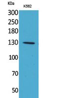
- 135kD
- 背景:
- This gene encodes a cytoplasmic protein that initiates apoptosis. This protein contains several copies of the WD-40 domain, a caspase recruitment domain (CARD), and an ATPase domain (NB-ARC). Upon binding cytochrome c and dATP, this protein forms an oligomeric apoptosome. The apoptosome binds and cleaves caspase 9 preproprotein, releasing its mature, activated form. Activated caspase 9 stimulates the subsequent caspase cascade that commits the cell to apoptosis. Alternative splicing results in several transcript variants encoding different isoforms. [provided by RefSeq, Jul 2008],
- 功能:
- domain:The CARD domain mediates interaction with APIP.,function:Oligomeric Apaf-1 mediates the cytochrome c-dependent autocatalytic activation of pro-caspase-9 (Apaf-3), leading to the activation of caspase-3 and apoptosis. This activation requires ATP. Isoform 6 is less effective in inducing apoptosis.,induction:By E2F and p53 in apoptotic neurons.,similarity:Contains 1 CARD domain.,similarity:Contains 1 NB-ARC domain.,similarity:Contains 13 WD repeats.,subunit:Monomer. Oligomerizes upon binding of cytochrome c and dATP. Oligomeric Apaf-1 and pro-caspase-9 bind to each other via their respective NH2-terminal CARD domains and consecutively mature caspase-9 is released from the complex. Pro-caspase-3 is recruited into the Apaf-1-pro-caspase-9 complex via interaction with pro-caspase-9. Interacts with APIP.,tissue specificity:Ubiquitous. Highest levels of expression in adult spleen and per
- 细胞定位:
- Cytoplasm .
- 组织表达:
- Ubiquitous. Highest levels of expression in adult spleen and peripheral blood leukocytes, and in fetal brain, kidney and lung. Isoform 1 is expressed in heart, kidney and liver.
Flavonoid-Rich Extract of Oldenlandia diffusa (Willd.) Roxb. Inhibits Gastric Cancer by Activation of Caspase-Dependent Mitochondrial Apoptosis Chinese Journal of Integrative Medicine Qiu-lan Wang WB Human
货号:YT5378
Cathepsin B mediates the lysosomal-mitochondrial apoptosis pathway in arsenic-induced microglial cell injury. Baofei Sun WB Mouse 1:1000 BV2 cell
货号:YT5378
- June 19-2018
- WESTERN IMMUNOBLOTTING PROTOCOL
- June 19-2018
- IMMUNOHISTOCHEMISTRY-PARAFFIN PROTOCOL
- June 19-2018
- IMMUNOFLUORESCENCE PROTOCOL
- September 08-2020
- FLOW-CYTOMEYRT-PROTOCOL
- May 20-2022
- Cell-Based ELISA│解您多样本WB检测之困扰
- July 13-2018
- CELL-BASED-ELISA-PROTOCOL-FOR-ACETYL-PROTEIN
- July 13-2018
- CELL-BASED-ELISA-PROTOCOL-FOR-PHOSPHO-PROTEIN
- July 13-2018
- Antibody-FAQs
- 产品图片

- Western Blot analysis of K562 cells using Apaf-1 Polyclonal Antibody. Secondary antibody(catalog#:RS0002) was diluted at 1:20000

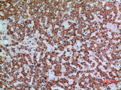
- Immunohistochemical analysis of paraffin-embedded human-liver, antibody was diluted at 1:100

- Immunohistochemical analysis of paraffin-embedded human-lung, antibody was diluted at 1:100

- Western blot analysis of lysate from K562 cells, using APAF1 Antibody.


