- 靶点:
- CD133
- 简介:
- >>Transcriptional misregulation in cancer
- 基因名称:
- PROM1
- 蛋白名称:
- Prominin-1
- Human Gene Id:
- 8842
- Human Swiss Prot No:
- O43490
- Mouse Swiss Prot No:
- O54990
- 免疫原:
- The antiserum was produced against synthesized peptide derived from the N-terminal region of human PROM1. AA range:41-90
- 特异性:
- CD133 Polyclonal Antibody detects endogenous levels of CD133 protein.
- 组成:
- Liquid in PBS containing 50% glycerol, 0.5% BSA and 0.02% sodium azide.
- 来源:
- Polyclonal, Rabbit,IgG
- 稀释:
- WB 1:500 - 1:2000. IHC: 1:100-300 ELISA: 1:20000. IF 1:100-300 Not yet tested in other applications.
- 纯化工艺:
- The antibody was affinity-purified from rabbit antiserum by affinity-chromatography using epitope-specific immunogen.
- 浓度:
- 1 mg/ml
- 储存:
- -15°C to -25°C/1 year(Do not lower than -25°C)
- 其他名称:
- PROM1;Prominin-1;Antigen AC133;Prominin-like protein 1;CD133
- 实测条带:
- 100kD
- 背景:
- This gene encodes a pentaspan transmembrane glycoprotein. The protein localizes to membrane protrusions and is often expressed on adult stem cells, where it is thought to function in maintaining stem cell properties by suppressing differentiation. Mutations in this gene have been shown to result in retinitis pigmentosa and Stargardt disease. Expression of this gene is also associated with several types of cancer. This gene is expressed from at least five alternative promoters that are expressed in a tissue-dependent manner. Multiple transcript variants encoding different isoforms have been found for this gene. [provided by RefSeq, Mar 2009],
- 功能:
- disease:Defects in PROM1 are the cause of cone-rod dystrophy type 12 (CORD12) [MIM:612657]. CORD12 is an inherited retinal dystrophy characterized by retinal pigment deposits visible on fundus examination, predominantly in the macular region, and initial loss of cone photoreceptors followed by rod degeneration. This leads to decreased visual acuity and sensitivity in the central visual field, followed by loss of peripheral vision. Severe loss of vision occurs earlier than in retinitis pigmentosa.,disease:Defects in PROM1 are the cause of retinal macular dystrophy type 2 (MCDR2) [MIM:608051]. MCDR2 is a bull's-eye macular dystrophy characterized by bilateral annular atrophy of retinal pigment epithelium at the macula.,disease:Defects in PROM1 are the cause of retinitis pigmentosa type 41 (RP41) [MIM:612095]; also known as retinal degeneration autosomal recessive prominin-related. RP is a
- 细胞定位:
- Apical cell membrane ; Multi-pass membrane protein . Cell projection, microvillus membrane ; Multi-pass membrane protein . Cell projection, cilium, photoreceptor outer segment . Endoplasmic reticulum. Endoplasmic reticulum-Golgi intermediate compartment. Found in extracellular membrane particles in various body fluids such as cerebrospinal fluid, saliva, seminal fluid and urine.
- 组织表达:
- Isoform 1 is selectively expressed on CD34 hematopoietic stem and progenitor cells in adult and fetal bone marrow, fetal liver, cord blood and adult peripheral blood. Isoform 1 is not detected on other blood cells. Isoform 1 is also expressed in a number of non-lymphoid tissues including retina, pancreas, placenta, kidney, liver, lung, brain and heart. Found in saliva within small membrane particles. Isoform 2 is predominantly expressed in fetal liver, skeletal muscle, kidney, and heart as well as adult pancreas, kidney, liver, lung, and placenta. Isoform 2 is highly expressed in fetal liver, low in bone marrow, and barely detectable in peripheral blood. Isoform 2 is expressed on hematopoietic stem cells and in epidermal basal cells (at protein level). Expressed in adult retina by rod and
Prolonged inhibition of class I PI3K promotes liver cancer stem cell expansion by augmenting SGK3/GSK-3β/β-catenin signalling. JOURNAL OF EXPERIMENTAL & CLINICAL CANCER RESEARCH 2018 Jun 25 WB Human MHCC-97H cell,Huh7 cell
货号:YT5192
Black TiO2-based nanoprobes for T1-weighted MRI-guided photothermal therapy in CD133 high expressed pancreatic cancer stem-like cells. Biomaterials Science Biomater Sci-Uk. 2018 Jul;6(8):2209-2218 WB Human 1 : 2000 PANC-1 cell,SW1990 cell
货号:YT5192
Secreted protein acidic and rich in cysteine promotes epithelial–mesenchymal transition of hepatocellular carcinoma cells and acquisition of cancerstem cell phenotypes. JOURNAL OF GASTROENTEROLOGY AND HEPATOLOGY J Gastroen Hepatol. 2019 Oct;34(10):1860-1868 WB Human 1:1000 MHCC-97H cell, Huh-7 cell, SMMC-7721 cell
货号:YT5192
Wang, Siqi, et al. "Black TiO2 based nanoprobes for T1-weighted MRI guided photothermal therapy in CD133 high expressed pancreatic cancer stem-like cells." Biomaterials Science (2018).
货号:YT5192
Jiang, Xin, et al. "SPARC promotes epithelial‐mesenchymal transition of hepatocellular carcinoma cells and acquisition of cancer stem cell phenotypes." Journal of gastroenterology and hepatology (2019).
货号:YT5192
Fluid shear stress induces cell migration via RhoA-YAP1-autophagy pathway in liver cancer stem cells Cell Adhesion & Migration Wenzhi Guo WB Human
货号:YT5192
FEN1 upregulation mediated by SUMO2 via antagonizing proteasomal degradation promotes hepatocellular carcinoma stemness Translational Oncology Zhenxiang Peng WB Human Huh7 cell,MHCC-97H cell
货号:YT5192
- June 19-2018
- WESTERN IMMUNOBLOTTING PROTOCOL
- June 19-2018
- IMMUNOHISTOCHEMISTRY-PARAFFIN PROTOCOL
- June 19-2018
- IMMUNOFLUORESCENCE PROTOCOL
- September 08-2020
- FLOW-CYTOMEYRT-PROTOCOL
- May 20-2022
- Cell-Based ELISA│解您多样本WB检测之困扰
- July 13-2018
- CELL-BASED-ELISA-PROTOCOL-FOR-ACETYL-PROTEIN
- July 13-2018
- CELL-BASED-ELISA-PROTOCOL-FOR-PHOSPHO-PROTEIN
- July 13-2018
- Antibody-FAQs
- 产品图片
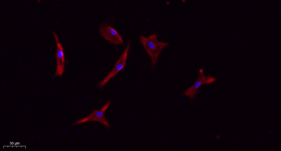
- Immunofluorescence analysis of A549. 1,primary Antibody(red) was diluted at 1:200(4°C overnight). 2, Goat Anti Rabbit IgG (H&L) - Alexa Fluor 594 Secondary antibody was diluted at 1:1000(room temperature, 50min).3, Picture B: DAPI(blue) 10min.

- Western Blot analysis of A549, SW620, HT29, Jurkat, mouse kidney, mouse lung cells using CD133 Polyclonal Antibody. Secondary antibody(catalog#:RS0002) was diluted at 1:20000
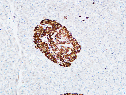
- Immunohistochemical analysis of paraffin-embedded Human pancreas. 1, Antibody was diluted at 1:200(4° overnight). 2, High-pressure and temperature EDTA, pH8.0 was used for antigen retrieval. 3,Secondary antibody was diluted at 1:200(room temperature, 30min).
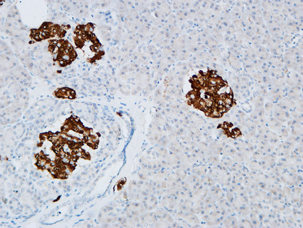
- Immunohistochemical analysis of paraffin-embedded Human pancreas. 1, Antibody was diluted at 1:200(4° overnight). 2, High-pressure and temperature EDTA, pH8.0 was used for antigen retrieval. 3,Secondary antibody was diluted at 1:200(room temperature, 30min).
- The picture was kindly provided by our customer

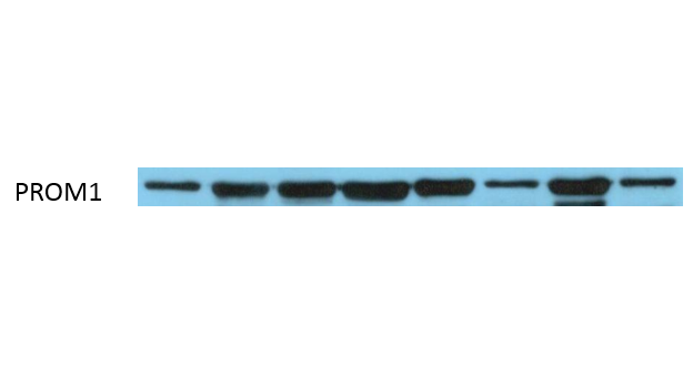
- Western Blot analysis of various cells. The picture was provided by our customer.


