- 靶点:
- DR3
- 简介:
- >>Cytokine-cytokine receptor interaction
- 基因名称:
- TNFRSF25
- 蛋白名称:
- Tumor necrosis factor receptor superfamily member 25
- Human Gene Id:
- 8718
- Human Swiss Prot No:
- Q93038
- 免疫原:
- Synthesized peptide derived from DR3 . at AA range: 230-310
- 特异性:
- DR3 Polyclonal Antibody detects endogenous levels of DR3 protein.
- 组成:
- Liquid in PBS containing 50% glycerol, 0.5% BSA and 0.02% sodium azide.
- 来源:
- Polyclonal, Rabbit,IgG
- 稀释:
- WB 1:500 - 1:2000. IHC: 1:100-300 ELISA: 1:20000.. IF 1:50-200
- 纯化工艺:
- The antibody was affinity-purified from rabbit antiserum by affinity-chromatography using epitope-specific immunogen.
- 浓度:
- 1 mg/ml
- 储存:
- -15°C to -25°C/1 year(Do not lower than -25°C)
- 其他名称:
- TNFRSF25;APO3;DDR3;DR3;TNFRSF12;WSL;WSL1;Tumor necrosis factor receptor superfamily member 25;Apo-3;Apoptosis-inducing receptor AIR;Apoptosis-mediating receptor DR3;Apoptosis-mediating receptor TRAMP;Death receptor 3;Lymphocyte-associated receptor of death;LARD;Protein WSL;Protein WSL-1
- 实测条带:
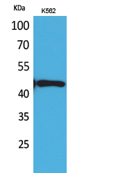
- 45kD
- 背景:
- The protein encoded by this gene is a member of the TNF-receptor superfamily. This receptor is expressed preferentially in the tissues enriched in lymphocytes, and it may play a role in regulating lymphocyte homeostasis. This receptor has been shown to stimulate NF-kappa B activity and regulate cell apoptosis. The signal transduction of this receptor is mediated by various death domain containing adaptor proteins. Knockout studies in mice suggested the role of this gene in the removal of self-reactive T cells in the thymus. Multiple alternatively spliced transcript variants of this gene encoding distinct isoforms have been reported, most of which are potentially secreted molecules. The alternative splicing of this gene in B and T cells encounters a programmed change upon T-cell activation, which predominantly produces full-length, membrane bound isoforms, and is thought to be involve
- 功能:
- function:Receptor for TNFSF12/APO3L/TWEAK. Interacts directly with the adapter TRADD. Mediates activation of NF-kappa-B and induces apoptosis. May play a role in regulating lymphocyte homeostasis.,PTM:Glycosylated.,similarity:Contains 1 death domain.,similarity:Contains 4 TNFR-Cys repeats.,subunit:Homodimer. Interacts strongly via the death domains with TNFRSF1 and TRADD to activate at least two distinct signaling cascades, apoptosis and NF-kappa-B signaling. Interacts with BAG4.,tissue specificity:Abundantly expressed in thymocytes and lymphocytes. Detected in lymphocyte-rich tissues such as thymus, colon, intestine, and spleen. Also found in the prostate.,
- 细胞定位:
- [Isoform 1]: Cell membrane; Single-pass type I membrane protein.; [Isoform 2]: Cell membrane; Single-pass type I membrane protein.; [Isoform 9]: Cell membrane; Single-pass type I membrane protein.; [Isoform 11]: Cell membrane; Single-pass type I membrane protein.; [Isoform 3]: Secreted.; [Isoform 4]: Secreted.; [Isoform 5]: Secreted.; [Isoform 6]: Secreted.; [Isoform 7]: Secreted.; [Isoform 8]: Secreted.; [Isoform 10]: Secreted.; [Isoform 12]: Secreted.
- 组织表达:
- Abundantly expressed in thymocytes and lymphocytes. Detected in lymphocyte-rich tissues such as thymus, colon, intestine, and spleen. Also found in the prostate.
Anti-TL1A monoclonal antibody modulates the dysregulation of Th1/Th17 cells and attenuates granuloma formation in sarcoidosis by inhibiting the PI3K/AKT signaling pathway INTERNATIONAL IMMUNOPHARMACOLOGY Chengxing Ma WB,IHC Mouse,Human lung tissue
货号:YT5135
- June 19-2018
- WESTERN IMMUNOBLOTTING PROTOCOL
- June 19-2018
- IMMUNOHISTOCHEMISTRY-PARAFFIN PROTOCOL
- June 19-2018
- IMMUNOFLUORESCENCE PROTOCOL
- September 08-2020
- FLOW-CYTOMEYRT-PROTOCOL
- May 20-2022
- Cell-Based ELISA│解您多样本WB检测之困扰
- July 13-2018
- CELL-BASED-ELISA-PROTOCOL-FOR-ACETYL-PROTEIN
- July 13-2018
- CELL-BASED-ELISA-PROTOCOL-FOR-PHOSPHO-PROTEIN
- July 13-2018
- Antibody-FAQs
- 产品图片

- Western Blot analysis of K562 cells using DR3 Polyclonal Antibody. Secondary antibody(catalog#:RS0002) was diluted at 1:20000

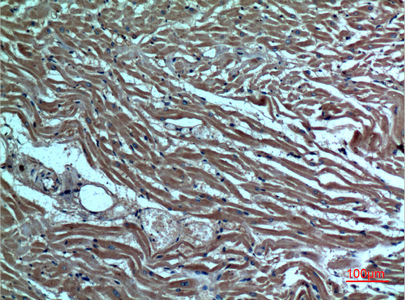
- Immunohistochemical analysis of paraffin-embedded human-heart, antibody was diluted at 1:100

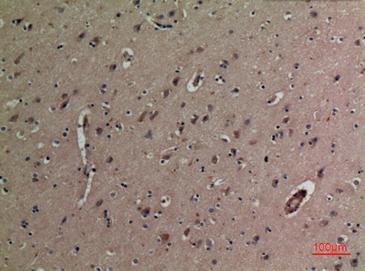
- Immunohistochemical analysis of paraffin-embedded human-brain, antibody was diluted at 1:100


