Tubulin α Polyclonal Antibody
- 货号:YT4777
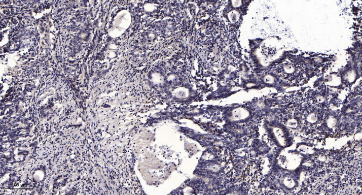
- 应用:WB;IHC;IF;ELISA
- 种属:Human;Mouse;Rat;Chicken
- 靶点:
- Tubulin α
- 简介:
- >>Phagosome;>>Apoptosis;>>Tight junction;>>Gap junction;>>Alzheimer disease;>>Parkinson disease;>>Amyotrophic lateral sclerosis;>>Huntington disease;>>Prion disease;>>Pathways of neurodegeneration - multiple diseases;>>Pathogenic Escherichia coli infection;>>Salmonella infection
- 基因名称:
- TUBA1A
- 蛋白名称:
- Tubulin alpha-1A chain
- Human Gene Id:
- 7846/10376
- Human Swiss Prot No:
- Q71U36/P68363
- Mouse Gene Id:
- 22142/22143
- Rat Gene Id:
- 64158/500929
- Rat Swiss Prot No:
- P68370/Q6P9V9
- 免疫原:
- The antiserum was produced against synthesized peptide derived from human Tubulin alpha. AA range:402-451
- 特异性:
- Tubulin α Polyclonal Antibody detects endogenous levels of Tubulin α protein.
- 组成:
- Liquid in PBS containing 50% glycerol, 0.5% BSA and 0.02% sodium azide.
- 来源:
- Polyclonal, Rabbit,IgG
- 稀释:
- WB 1:500 - 1:2000. IHC 1:100 - 1:300. IF 1:200 - 1:1000. ELISA: 1:5000. Not yet tested in other applications.
- 纯化工艺:
- The antibody was affinity-purified from rabbit antiserum by affinity-chromatography using epitope-specific immunogen.
- 浓度:
- 1 mg/ml
- 储存:
- -15°C to -25°C/1 year(Do not lower than -25°C)
- 其他名称:
- TUBA1A;TUBA3;Tubulin alpha-1A chain;Alpha-tubulin 3;Tubulin B-alpha-1;Tubulin alpha-3 chain;TUBA1B;Tubulin alpha-1B chain;Alpha-tubulin ubiquitous;Tubulin K-alpha-1;Tubulin alpha-ubiquitous chain
- 实测条带:
- 50kD
- 背景:
- Microtubules of the eukaryotic cytoskeleton perform essential and diverse functions and are composed of a heterodimer of alpha and beta tubulins. The genes encoding these microtubule constituents belong to the tubulin superfamily, which is composed of six distinct families. Genes from the alpha, beta and gamma tubulin families are found in all eukaryotes. The alpha and beta tubulins represent the major components of microtubules, while gamma tubulin plays a critical role in the nucleation of microtubule assembly. There are multiple alpha and beta tubulin genes, which are highly conserved among species. This gene encodes alpha tubulin and is highly similar to the mouse and rat Tuba1 genes. Northern blotting studies have shown that the gene expression is predominantly found in morphologically differentiated neurologic cells. This gene is one of three alpha-tubulin genes in a cluster on chromosome 12q.
- 功能:
- disease:Defects in TUBA1A are the cause of lissencephaly type 3 (LIS3) [MIM:611603]. LIS is characterized by a smooth brain surface due to the absence (agyria) or reduction (pachygyria) of surface convolutions. It is often associated with psychomotor retardation and seizures. LIS3 features include agyria or pachygyria or laminar heterotopia, severe mental retardation, motor delay, variable presence of seizures, and abnormalities of corpus callosum, hippocampus, cerebellar vermis and brainstem.,function:Tubulin is the major constituent of microtubules. It binds two moles of GTP, one at an exchangeable site on the beta chain and one at a non-exchangeable site on the alpha-chain.,PTM:Undergoes a tyrosination/detyrosination cycle, the cyclic removal and re-addition of a C-terminal tyrosine residue by the enzymes tubulin tyrosine carboxypeptidase (TTCP) and tubulin tyrosine ligase (TTL), resp
- 细胞定位:
- Cytoplasm, cytoskeleton.
- 组织表达:
- Expressed at a high level in fetal brain.
- June 19-2018
- WESTERN IMMUNOBLOTTING PROTOCOL
- June 19-2018
- IMMUNOHISTOCHEMISTRY-PARAFFIN PROTOCOL
- June 19-2018
- IMMUNOFLUORESCENCE PROTOCOL
- September 08-2020
- FLOW-CYTOMEYRT-PROTOCOL
- May 20-2022
- Cell-Based ELISA│解您多样本WB检测之困扰
- July 13-2018
- CELL-BASED-ELISA-PROTOCOL-FOR-ACETYL-PROTEIN
- July 13-2018
- CELL-BASED-ELISA-PROTOCOL-FOR-PHOSPHO-PROTEIN
- July 13-2018
- Antibody-FAQs