Smad2 Polyclonal Antibody

    • 货号:YT4329
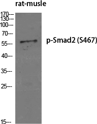
    • 应用:WB;CoIP;IHC;IF;ELISA
    • 种属:Human;Mouse;Rat
      • 靶点:
      • Smad2
      • 简介:
      • >>Cell cycle;>>Endocytosis;>>Cellular senescence;>>TGF-beta signaling pathway;>>Apelin signaling pathway;>>Hippo signaling pathway;>>Signaling pathways regulating pluripotency of stem cells;>>Th17 cell differentiation;>>Relaxin signaling pathway;>>AGE-RAGE signaling pathway in diabetic complications;>>Chagas disease;>>Human T-cell leukemia virus 1 infection;>>Pathways in cancer;>>Proteoglycans in cancer;>>Colorectal cancer;>>Pancreatic cancer;>>Hepatocellular carcinoma;>>Gastric cancer;>>Inflammatory bowel disease;>>Diabetic cardiomyopathy
      • 基因名称:
      • SMAD2
      • 蛋白名称:
      • Mothers against decapentaplegic homolog 2
      • Human Gene Id:
      • 4087
      • Human Swiss Prot No:
      • Q15796
      • Mouse Swiss Prot No:
      • Q62432
      • 免疫原:
      • The antiserum was produced against synthesized peptide derived from human Smad2. AA range:418-467
      • 特异性:
      • Smad2 Polyclonal Antibody detects endogenous levels of Smad2 protein.
      • 组成:
      • Liquid in PBS containing 50% glycerol, 0.5% BSA and 0.02% sodium azide.
      • 来源:
      • Polyclonal, Rabbit,IgG
      • 稀释:
      • WB 1:500 - 1:2000. IHC 1:100 - 1:300. IF 1:200 - 1:1000. ELISA: 1:5000. Not yet tested in other applications.
      • 纯化工艺:
      • The antibody was affinity-purified from rabbit antiserum by affinity-chromatography using epitope-specific immunogen.
      • 浓度:
      • 1 mg/ml
      • 储存:
      • -15°C to -25°C/1 year(Do not lower than -25°C)
      • 其他名称:
      • SMAD2;MADH2;MADR2;Mothers against decapentaplegic homolog 2;MAD homolog 2;Mothers against DPP homolog 2;JV18-1;Mad-related protein 2;hMAD-2;SMAD family member 2;SMAD 2;Smad2;hSMAD2
      • 实测条带:
      • 58kD
      • 背景:
      • The protein encoded by this gene belongs to the SMAD, a family of proteins similar to the gene products of the Drosophila gene 'mothers against decapentaplegic' (Mad) and the C. elegans gene Sma. SMAD proteins are signal transducers and transcriptional modulators that mediate multiple signaling pathways. This protein mediates the signal of the transforming growth factor (TGF)-beta, and thus regulates multiple cellular processes, such as cell proliferation, apoptosis, and differentiation. This protein is recruited to the TGF-beta receptors through its interaction with the SMAD anchor for receptor activation (SARA) protein. In response to TGF-beta signal, this protein is phosphorylated by the TGF-beta receptors. The phosphorylation induces the dissociation of this protein with SARA and the association with the family member SMAD4. The association with SMAD4 is important for the translocation
      • 功能:
      • disease:Defects in SMAD2 are found in sporadic cases of colorectal carcinoma.,function:Transcriptional modulator activated by TGF-beta and activin type 1 receptor kinase. SMAD2 is a receptor-regulated SMAD (R-SMAD). May act as a tumor suppressor in colorectal carcinoma.,PTM:Acetylated on Lys-19 by coactivators in response to TGF-beta signaling, which increases transcriptional activity. Isoform short: Acetylation increases DNA binding activity in vitro and enhances its association with target promoters in vivo.,PTM:In response to TGF-beta, ubiquitinated by NEDD4L; which promotes its degradation.,PTM:Phosphorylated on one or several of Thr-220, Ser-245, Ser-250, and Ser-255. In response to TGF-beta, phosphorylated on Ser-465/467 by TGF-beta and activin type 1 receptor kinases. Able to interact with SMURF2 when phosphorylated on Ser-465/467, recruiting other proteins, such as SNON, for degr
      • 细胞定位:
      • Cytoplasm . Nucleus . Cytoplasmic and nuclear in the absence of TGF-beta. On TGF-beta stimulation, migrates to the nucleus when complexed with SMAD4 (PubMed:9865696, PubMed:21145499). On dephosphorylation by phosphatase PPM1A, released from the SMAD2/SMAD4 complex, and exported out of the nucleus by interaction with RANBP1 (PubMed:16751101, PubMed:19289081). Localized mainly to the nucleus in the early stages of embryo development with expression becoming evident in the cytoplasm at the blastocyst and epiblast stages (By similarity). .
      • 组织表达:
      • Expressed at high levels in skeletal muscle, endothelial cells, heart and placenta.

      Human Novel MicroRNA Seq-915_x4024 in Keratinocytes Contributes to Skin Regeneration by Suppressing Scar Formation. Molecular Therapy-Nucleic Acids Mol Ther-Nucl Acids. 2019 Mar;14:410 WB Human HaCaT cell
      货号:YT4329

      Proteomic Profiling of Radiation-Induced Skin Fibrosis in Rats: Targeting the Ubiquitin-Proteasome System. INTERNATIONAL JOURNAL OF RADIATION ONCOLOGY BIOLOGY PHYSICS 2016 Jan 20 WB,IF Human HaCaT cell, WS-1 cell
      货号:YT4329

      Positive feedback loop of YB-1 interacting with Smad2 promotes liver fibrosis. BIOCHEMICAL AND BIOPHYSICAL RESEARCH COMMUNICATIONS Biochem Bioph Res Co. 2017 Mar;484:753 CoIP Human LX-2 cell,293T cell
      货号:YT4329

      Song, Jianyuan, et al. "The Role of FABP5 in Radiation-Induced Human Skin Fibrosis." Radiation research 189.2 (2017): 177-186.
      货号:YT4329

      Wang, Wenjie, et al. "Proteomic profiling of radiation-induced skin fibrosis in rats: Targeting the ubiquitin-proteasome system." International Journal of Radiation Oncology* Biology* Physics 95.2 (2016): 751-760.
      货号:YT4329

      Shao, Min, et al. "Exogenous angiotensin (1-7) directly inhibits epithelial-mesenchymal transformation induced by transforming growth factor-β1 in alveolar epithelial cells." Biomedicine & Pharmacotherapy 117 (2019): 109193.
      货号:YT4329

      • 产品图片
      • Immunofluorescence analysis of Hela cell. 1,Smad2 Polyclonal Antibody(red) was diluted at 1:200(4° overnight). MICU1 Monoclonal Antibody(Mix)(green) was diluted at 1:200(4° overnight). 2, Goat Anti Rabbit Alexa Fluor 594 Catalog:RS3611 was diluted at 1:1000(room temperature, 50min). Goat Anti Mouse Alexa Fluor 488 Catalog:RS3208 was diluted at 1:1000(room temperature, 50min).
      • Western blot analysis of Smad2 Polyclonal Antibody, using Hela, HepG2,SW480, HEK293 cell, 4° over night, secondary antibody(cat: RS0002 was diluted at 1:10000, 37° 1hour.
      • Immunofluorescence analysis of rat-lung tissue. 1,Smad2 Polyclonal Antibody(red) was diluted at 1:200(4°C,overnight). 2, Cy3 labled Secondary antibody was diluted at 1:300(room temperature, 50min).3, Picture B: DAPI(blue) 10min. Picture A:Target. Picture B: DAPI. Picture C: merge of A+B
      • Immunofluorescence analysis of rat-lung tissue. 1,Smad2 Polyclonal Antibody(red) was diluted at 1:200(4°C,overnight). 2, Cy3 labled Secondary antibody was diluted at 1:300(room temperature, 50min).3, Picture B: DAPI(blue) 10min. Picture A:Target. Picture B: DAPI. Picture C: merge of A+B
      • Immunofluorescence analysis of rat-spleen tissue. 1,Smad2 Polyclonal Antibody(red) was diluted at 1:200(4°C,overnight). 2, Cy3 labled Secondary antibody was diluted at 1:300(room temperature, 50min).3, Picture B: DAPI(blue) 10min. Picture A:Target. Picture B: DAPI. Picture C: merge of A+B
      • Immunofluorescence analysis of rat-spleen tissue. 1,Smad2 Polyclonal Antibody(red) was diluted at 1:200(4°C,overnight). 2, Cy3 labled Secondary antibody was diluted at 1:300(room temperature, 50min).3, Picture B: DAPI(blue) 10min. Picture A:Target. Picture B: DAPI. Picture C: merge of A+B
      • Immunofluorescence analysis of mouse-lung tissue. 1,Smad2 Polyclonal Antibody(red) was diluted at 1:200(4°C,overnight). 2, Cy3 labled Secondary antibody was diluted at 1:300(room temperature, 50min).3, Picture B: DAPI(blue) 10min. Picture A:Target. Picture B: DAPI. Picture C: merge of A+B
      • Immunofluorescence analysis of mouse-lung tissue. 1,Smad2 Polyclonal Antibody(red) was diluted at 1:200(4°C,overnight). 2, Cy3 labled Secondary antibody was diluted at 1:300(room temperature, 50min).3, Picture B: DAPI(blue) 10min. Picture A:Target. Picture B: DAPI. Picture C: merge of A+B
      • Immunohistochemical analysis of paraffin-embedded Human-uterus tissue. 1,Smad2 Polyclonal Antibody was diluted at 1:200(4°C,overnight). 2, Sodium citrate pH 6.0 was used for antibody retrieval(>98°C,20min). 3,Secondary antibody was diluted at 1:200(room tempeRature, 30min). Negative control was used by secondary antibody only.
      • Immunohistochemical analysis of paraffin-embedded Human-uterus-cancer tissue. 1,Smad2 Polyclonal Antibody was diluted at 1:200(4°C,overnight). 2, Sodium citrate pH 6.0 was used for antibody retrieval(>98°C,20min). 3,Secondary antibody was diluted at 1:200(room tempeRature, 30min). Negative control was used by secondary antibody only.
      • Immunohistochemical analysis of paraffin-embedded Human-stomach tissue. 1,Smad2 Polyclonal Antibody was diluted at 1:200(4°C,overnight). 2, Sodium citrate pH 6.0 was used for antibody retrieval(>98°C,20min). 3,Secondary antibody was diluted at 1:200(room tempeRature, 30min). Negative control was used by secondary antibody only.
      • Immunohistochemical analysis of paraffin-embedded Human-stomach-cancer tissue. 1,Smad2 Polyclonal Antibody was diluted at 1:200(4°C,overnight). 2, Sodium citrate pH 6.0 was used for antibody retrieval(>98°C,20min). 3,Secondary antibody was diluted at 1:200(room tempeRature, 30min). Negative control was used by secondary antibody only.
      • Immunohistochemical analysis of paraffin-embedded Rat-spinal-cord tissue. 1,Smad2 Polyclonal Antibody was diluted at 1:200(4°C,overnight). 2, Sodium citrate pH 6.0 was used for antibody retrieval(>98°C,20min). 3,Secondary antibody was diluted at 1:200(room tempeRature, 30min). Negative control was used by secondary antibody only.
      • Immunohistochemical analysis of paraffin-embedded Rat-brain tissue. 1,Smad2 Polyclonal Antibody was diluted at 1:200(4°C,overnight). 2, Sodium citrate pH 6.0 was used for antibody retrieval(>98°C,20min). 3,Secondary antibody was diluted at 1:200(room tempeRature, 30min). Negative control was used by secondary antibody only.
      • Immunohistochemical analysis of paraffin-embedded Mouse-testis tissue. 1,Smad2 Polyclonal Antibody was diluted at 1:200(4°C,overnight). 2, Sodium citrate pH 6.0 was used for antibody retrieval(>98°C,20min). 3,Secondary antibody was diluted at 1:200(room tempeRature, 30min). Negative control was used by secondary antibody only.
      • Immunohistochemical analysis of paraffin-embedded Mouse-colon tissue. 1,Smad2 Polyclonal Antibody was diluted at 1:200(4°C,overnight). 2, Sodium citrate pH 6.0 was used for antibody retrieval(>98°C,20min). 3,Secondary antibody was diluted at 1:200(room tempeRature, 30min). Negative control was used by secondary antibody only.
      • Immunohistochemical analysis of paraffin-embedded Mouse-kidney tissue. 1,Smad2 Polyclonal Antibody was diluted at 1:200(4°C,overnight). 2, Sodium citrate pH 6.0 was used for antibody retrieval(>98°C,20min). 3,Secondary antibody was diluted at 1:200(room tempeRature, 30min). Negative control was used by secondary antibody only.
      • Immunohistochemical analysis of paraffin-embedded Mouse-brain tissue. 1,Smad2 Polyclonal Antibody was diluted at 1:200(4°C,overnight). 2, Sodium citrate pH 6.0 was used for antibody retrieval(>98°C,20min). 3,Secondary antibody was diluted at 1:200(room tempeRature, 30min). Negative control was used by secondary antibody only.
      • Immunohistochemical analysis of paraffin-embedded Mouse-spleen tissue. 1,Smad2 Polyclonal Antibody was diluted at 1:200(4°C,overnight). 2, Sodium citrate pH 6.0 was used for antibody retrieval(>98°C,20min). 3,Secondary antibody was diluted at 1:200(room tempeRature, 30min). Negative control was used by secondary antibody only.
      • Western Blot analysis of COLO cells using Smad2 Polyclonal Antibody diluted at 1:1000
      • Western blot analysis of KB lysis using Smad2 antibody. Antibody was diluted at 1:1000
      • Immunofluorescence analysis of HepG2 cells, using Smad2 Antibody. The picture on the right is blocked with the synthesized peptide.
      • Immunohistochemistry analysis of paraffin-embedded human prostate carcinoma tissue, using Smad2 Antibody. The picture on the right is blocked with the synthesized peptide.
      • Western blot analysis of lysates from HepG2 cells, using Smad2 Antibody. The lane on the right is blocked with the synthesized peptide.