- 首页
- 公司介绍
- 热门促销
-
全部产品
-
试剂盒
- |
-
一抗
- |
-
二抗
- |
-
蛋白
- |
-
免疫组化试剂
- |
-
WB 试剂
- PonceauS Staining Solution
- PBST Washing Buffer, 10X
- 1.5M Tris-HCl Buffer, pH8.8
- 1M Tris-HCl Buffer, pH6.8
- 10% SDS Solution
- Prestained Protein Marker
- TBST Washing Buffer, 10X
- SDS PAGE Loading Buffer, 5X
- Stripping Buffered Solution
- Tris Buffer, pH7.4, 10X
- Total Protein Extraction Kit
- Running Buffer, 10X
- Transfer Buffer, 10X
- 30% Acr-Bis(29:1) Solution
- Tris电泳液速溶颗粒
- PBS(1X, premixed powder)
- TBS(1X, premixed powder)
- 快速封闭液
- 转膜液速溶颗粒
- Chemical reagents
- 公司新闻
- 营销网络
- 资源中心
- 联系我们
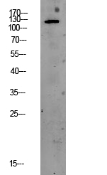
PERK Polyclonal Antibody
- 货号:YT3666
- 应用:IF;WB;IHC;ELISA
- 种属:Human;Mouse;Rat
- 简介:
- >>Mitophagy - animal;>>Autophagy - animal;>>Protein processing in endoplasmic reticulum;>>Apoptosis;>>Non-alcoholic fatty liver disease;>>Alzheimer disease;>>Parkinson disease;>>Amyotrophic lateral sclerosis;>>Prion disease;>>Pathways of neurodegeneration - multiple diseases;>>Hepatitis C;>>Measles;>>Herpes simplex virus 1 infection;>>Lipid and atherosclerosis
- 蛋白名称:
- Eukaryotic translation initiation factor 2-alpha kinase 3
- 免疫原:
- The antiserum was produced against synthesized peptide derived from human EIF2AK3. AA range:947-996
- 特异性:
- PERK Polyclonal Antibody detects endogenous levels of PERK protein.
- 组成:
- Liquid in PBS containing 50% glycerol, 0.5% BSA and 0.02% sodium azide.
- 来源:
- Polyclonal, Rabbit,IgG
- 稀释:
- IF 1:50-200 WB 1:500 - 1:2000. IHC 1:100 - 1:300. ELISA: 1:40000. Not yet tested in other applications.
- 纯化工艺:
- The antibody was affinity-purified from rabbit antiserum by affinity-chromatography using epitope-specific immunogen.
- 储存:
- -15°C to -25°C/1 year(Do not lower than -25°C)
- 其他名称:
- EIF2AK3;PEK;PERK;Eukaryotic translation initiation factor 2-alpha kinase 3;PRKR-like endoplasmic reticulum kinase;Pancreatic eIF2-alpha kinase;HsPEK
- 背景:
- The protein encoded by this gene phosphorylates the alpha subunit of eukaryotic translation-initiation factor 2, leading to its inactivation, and thus to a rapid reduction of translational initiation and repression of global protein synthesis. This protein is thought to modulate mitochondrial function. It is a type I membrane protein located in the endoplasmic reticulum (ER), where it is induced by ER stress caused by malfolded proteins. Mutations in this gene are associated with Wolcott-Rallison syndrome. [provided by RefSeq, Sep 2015],
- 功能:
- catalytic activity:ATP + a protein = ADP + a phosphoprotein.,disease:Defects in EIF2AK3 are the cause of Wolcott-Rallison syndrome (WRS) [MIM:226980]; also known as multiple epiphyseal dysplasia with early-onset diabetes mellitus. WRS is a rare autosomal recessive disorder, characterized by permanent neonatal or early infancy insulin-dependent diabetes and, at a later age, epiphyseal dysplasia, osteoporosis, growth retardation and other multisystem manifestations, such as hepatic and renal dysfunctions, mental retardation and cardiovascular abnormalities.,domain:The lumenal domain senses perturbations in protein folding in the ER, probably through reversible interaction with HSPA5/BIP.,enzyme regulation:Perturbation in protein folding in the endoplasmic reticulum (ER) promotes reversible dissociation from HSPA5/BIP and oligomerization, resulting in transautophosphorylation and kinase act
- 细胞定位:
- Endoplasmic reticulum membrane; Single-pass type I membrane protein.
- 组织表达:
- Ubiquitous. A high level expression is seen in secretory tissues.
.jpg)
