- 首页
- 公司介绍
- 热门促销
-
全部产品
-
试剂盒
- |
-
一抗
- |
-
二抗
- |
-
蛋白
- |
-
免疫组化试剂
- |
-
WB 试剂
- PonceauS Staining Solution
- PBST Washing Buffer, 10X
- 1.5M Tris-HCl Buffer, pH8.8
- 1M Tris-HCl Buffer, pH6.8
- 10% SDS Solution
- Prestained Protein Marker
- TBST Washing Buffer, 10X
- SDS PAGE Loading Buffer, 5X
- Stripping Buffered Solution
- Tris Buffer, pH7.4, 10X
- Total Protein Extraction Kit
- Running Buffer, 10X
- Transfer Buffer, 10X
- 30% Acr-Bis(29:1) Solution
- Tris电泳液速溶颗粒
- PBS(1X, premixed powder)
- TBS(1X, premixed powder)
- 快速封闭液
- 转膜液速溶颗粒
- Chemical reagents
- 公司新闻
- 营销网络
- 资源中心
- 联系我们
Myp Polyclonal Antibody
- 货号:YT2953
- 应用:WB;IHC;IF;ELISA
- 种属:Human;Rat;Mouse;
- 蛋白名称:
- Nucleolar protein 3
- 免疫原:
- The antiserum was produced against synthesized peptide derived from human ARC. AA range:159-208
- 特异性:
- Myp Polyclonal Antibody detects endogenous levels of Myp protein.
- 组成:
- Liquid in PBS containing 50% glycerol, 0.5% BSA and 0.02% sodium azide.
- 来源:
- Polyclonal, Rabbit,IgG
- 稀释:
- WB 1:500 - 1:2000. IHC 1:100 - 1:300. IF 1:200 - 1:1000. ELISA: 1:20000. Not yet tested in other applications.
- 纯化工艺:
- The antibody was affinity-purified from rabbit antiserum by affinity-chromatography using epitope-specific immunogen.
- 储存:
- -15°C to -25°C/1 year(Do not lower than -25°C)
- 其他名称:
- Myp;NOL3;Nop30;Nucleolar protein 3;apoptosis repressor ARC;apoptosis repressor with CARD;apoptosis repressor with caspase recruitment domain (CARD);muscle-enriched cytoplasmic protein;nucleolar protein of 30 kDa
- 背景:
- NOL3 encodes an anti-apoptotic protein nucleolar protein 3 that has been shown to down-regulate the enzyme activities of caspase 2, caspase 8 and tumor protein p53. Multiple transcript variants encoding different isoforms have been found for NOL3.
- 细胞定位:
- [Isoform 1]: Nucleus, nucleolus . The SR-rich C-terminus mediates nuclear localization. .; [Isoform 3]: Cytoplasm .; [Isoform 2]: Cytoplasm . Mitochondrion . Sarcoplasmic reticulum . Membrane ; Lipid-anchor . Phosphorylation at Thr-149 results in translocation to mitochondria. Colocalized with mitochondria in response to oxidative stress. .
- 组织表达:
- Highly expressed in heart and skeletal muscle. Detected at low levels in placenta, liver, kidney and pancreas.

- Western Blot analysis of various cells using Myp Polyclonal Antibody
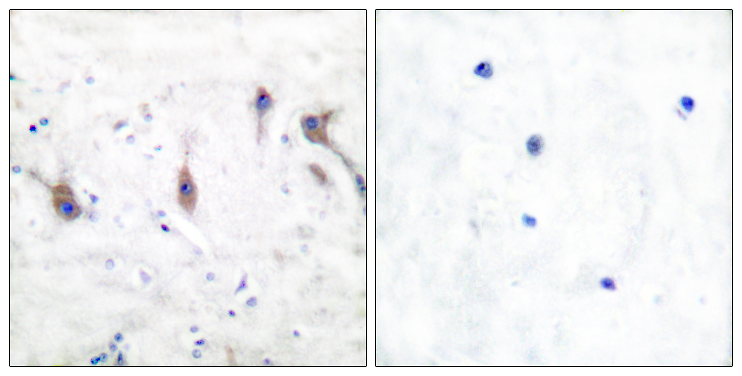
- Immunohistochemistry analysis of paraffin-embedded human brain tissue, using ARC Antibody. The picture on the right is blocked with the synthesized peptide.
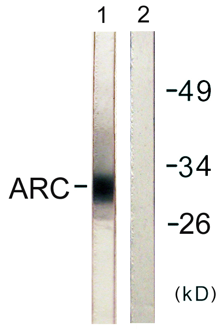
- Western blot analysis of lysates from HeLa cells, using ARC Antibody. The lane on the right is blocked with the synthesized peptide.

- Western Blot analysis of lane1 mouse-brain, lane2 mouse-kidney, lane3 Hela. land4 MCF7, lane5 293T, lane6 mouse-muscle using primary antibody at 1:1000 dilution 4°C, overnight. Secondary antibody(catalog#:RS23920) was diluted at 1:10000 25°C,1.5hours


