- 靶点:
- JAK1
- 简介:
- >>EGFR tyrosine kinase inhibitor resistance;>>PI3K-Akt signaling pathway;>>Necroptosis;>>Osteoclast differentiation;>>Signaling pathways regulating pluripotency of stem cells;>>NOD-like receptor signaling pathway;>>JAK-STAT signaling pathway;>>Th1 and Th2 cell differentiation;>>Th17 cell differentiation;>>Leishmaniasis;>>Toxoplasmosis;>>Tuberculosis;>>Hepatitis C;>>Hepatitis B;>>Measles;>>Human cytomegalovirus infection;>>Influenza A;>>Human papillomavirus infection;>>Human T-cell leukemia virus 1 infection;>>Kaposi sarcoma-associated herpesvirus infection;>>Herpes simplex virus 1 infection;>>Epstein-Barr virus infection;>>Coronavirus disease - COVID-19;>>Pathways in cancer;>>Viral carcinogenesis;>>Pancreatic cancer;>>PD-L1 expression and PD-1 checkpoint pathway in cancer
- 基因名称:
- JAK1
- 蛋白名称:
- Tyrosine-protein kinase JAK1
- Human Gene Id:
- 3716
- Human Swiss Prot No:
- P23458
- Mouse Swiss Prot No:
- P52332
- 免疫原:
- The antiserum was produced against synthesized peptide derived from human JAK1. AA range:988-1037
- 特异性:
- JAK1 Polyclonal Antibody detects endogenous levels of JAK1 protein.
- 组成:
- Liquid in PBS containing 50% glycerol, 0.5% BSA and 0.02% sodium azide.
- 来源:
- Polyclonal, Rabbit,IgG
- 稀释:
- IF 1:50-200 WB 1:200 - 1:1000. IHC 1:100 - 1:300. ELISA: 1:10000. Not yet tested in other applications.
- 纯化工艺:
- The antibody was affinity-purified from rabbit antiserum by affinity-chromatography using epitope-specific immunogen.
- 浓度:
- 1 mg/ml
- 储存:
- -15°C to -25°C/1 year(Do not lower than -25°C)
- 其他名称:
- JAK1;JAK1A;JAK1B;Tyrosine-protein kinase JAK1;Janus kinase 1;JAK-1
- 实测条带:
- 132kD
- 背景:
- This gene encodes a membrane protein that is a member of a class of protein-tyrosine kinases (PTK) characterized by the presence of a second phosphotransferase-related domain immediately N-terminal to the PTK domain. The encoded kinase phosphorylates STAT proteins (signal transducers and activators of transcription) and plays a key role in interferon-alpha/beta and interferon-gamma signal transduction. Alternative splicing results in multiple transcript variants. [provided by RefSeq, Mar 2016],
- 功能:
- catalytic activity:ATP + a [protein]-L-tyrosine = ADP + a [protein]-L-tyrosine phosphate.,domain:Possesses two phosphotransferase domains. The second one probably contains the catalytic domain (By similarity), while the presence of slight differences suggest a different role for domain 1.,domain:The FERM domain mediates interaction with JAKMIP1.,function:Tyrosine kinase of the non-receptor type, involved in the IFN-alpha/beta/gamma signal pathway. Kinase partner for the interleukin (IL)-2 receptor.,sequence caution:Translation N-terminally extended.,similarity:Belongs to the protein kinase superfamily. Tyr protein kinase family. JAK subfamily.,similarity:Contains 1 FERM domain.,similarity:Contains 1 protein kinase domain.,similarity:Contains 1 SH2 domain.,subcellular location:Wholly intracellular, possibly membrane associated.,subunit:Interacts with IL31RA, JAKMIP1 and SHB.,tissue specif
- 细胞定位:
- Endomembrane system; Peripheral membrane protein. Wholly intracellular, possibly membrane associated.
- 组织表达:
- Expressed at higher levels in primary colon tumors than in normal colon tissue. The expression level in metastatic colon tumors is comparable to the expression level in normal colon tissue.
Alpha-Momorcharin Inhibits Proinflammatory Cytokine Expression by M1 Macrophages but Not Anti-Inflammatory Cytokine Expression by M2 Macrophages Journal of Inflammation Research Fubing Shen WB Human
货号:YT2424
Phillygenin inhibited M1 macrophage polarization and reduced hepatic stellate cell activation by inhibiting macrophage exosomal miR-125b-5p BIOMEDICINE & PHARMACOTHERAPY Cheng Ma WB Mouse RAW264.7 cells
货号:YT2424
- June 19-2018
- WESTERN IMMUNOBLOTTING PROTOCOL
- June 19-2018
- IMMUNOHISTOCHEMISTRY-PARAFFIN PROTOCOL
- June 19-2018
- IMMUNOFLUORESCENCE PROTOCOL
- September 08-2020
- FLOW-CYTOMEYRT-PROTOCOL
- May 20-2022
- Cell-Based ELISA│解您多样本WB检测之困扰
- July 13-2018
- CELL-BASED-ELISA-PROTOCOL-FOR-ACETYL-PROTEIN
- July 13-2018
- CELL-BASED-ELISA-PROTOCOL-FOR-PHOSPHO-PROTEIN
- July 13-2018
- Antibody-FAQs
- 产品图片
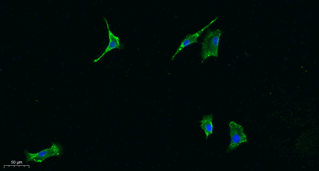
- Immunofluorescence analysis of A549. 1,primary Antibody was diluted at 1:200(4°C overnight). 2, Goat Anti Rabbit IgG (H&L) - Alexa Fluor 488 Secondary antibody was diluted at 1:1000(room temperature, 50min).3, Picture B: DAPI(blue) 10min.
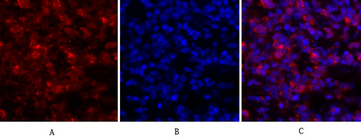
- Immunofluorescence analysis of rat-lung tissue. 1,JAK1 Polyclonal Antibody(red) was diluted at 1:200(4°C,overnight). 2, Cy3 labled Secondary antibody was diluted at 1:300(room temperature, 50min).3, Picture B: DAPI(blue) 10min. Picture A:Target. Picture B: DAPI. Picture C: merge of A+B
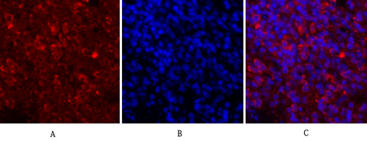
- Immunofluorescence analysis of rat-spleen tissue. 1,JAK1 Polyclonal Antibody(red) was diluted at 1:200(4°C,overnight). 2, Cy3 labled Secondary antibody was diluted at 1:300(room temperature, 50min).3, Picture B: DAPI(blue) 10min. Picture A:Target. Picture B: DAPI. Picture C: merge of A+B
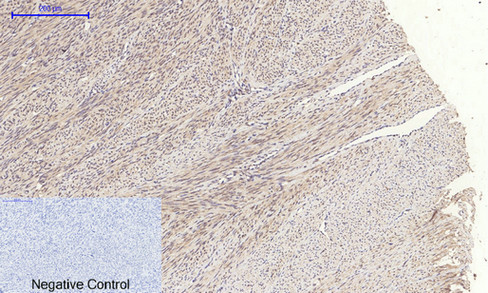
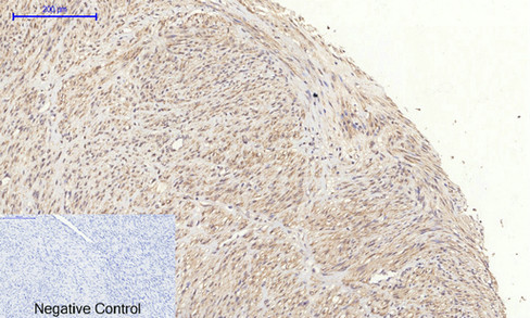
- Immunohistochemical analysis of paraffin-embedded Human-uterus tissue. 1,JAK1 Polyclonal Antibody was diluted at 1:200(4°C,overnight). 2, Sodium citrate pH 6.0 was used for antibody retrieval(>98°C,20min). 3,Secondary antibody was diluted at 1:200(room tempeRature, 30min). Negative control was used by secondary antibody only.
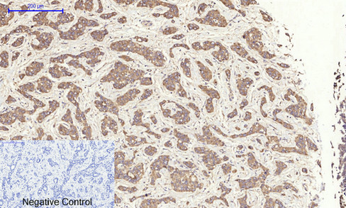
- Immunohistochemical analysis of paraffin-embedded Human-uterus-cancer tissue. 1,JAK1 Polyclonal Antibody was diluted at 1:200(4°C,overnight). 2, Sodium citrate pH 6.0 was used for antibody retrieval(>98°C,20min). 3,Secondary antibody was diluted at 1:200(room tempeRature, 30min). Negative control was used by secondary antibody only.
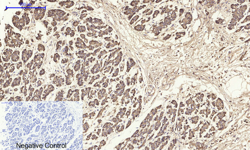
- Immunohistochemical analysis of paraffin-embedded Human-liver-cancer tissue. 1,JAK1 Polyclonal Antibody was diluted at 1:200(4°C,overnight). 2, Sodium citrate pH 6.0 was used for antibody retrieval(>98°C,20min). 3,Secondary antibody was diluted at 1:200(room tempeRature, 30min). Negative control was used by secondary antibody only.
- Immunohistochemical analysis of paraffin-embedded Human-stomach-cancer tissue. 1,JAK1 Polyclonal Antibody was diluted at 1:200(4°C,overnight). 2, Sodium citrate pH 6.0 was used for antibody retrieval(>98°C,20min). 3,Secondary antibody was diluted at 1:200(room tempeRature, 30min). Negative control was used by secondary antibody only.
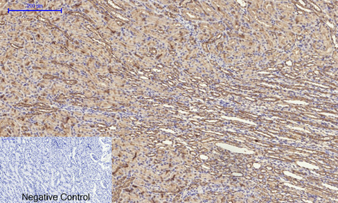
- Immunohistochemical analysis of paraffin-embedded Rat-kidney tissue. 1,JAK1 Polyclonal Antibody was diluted at 1:200(4°C,overnight). 2, Sodium citrate pH 6.0 was used for antibody retrieval(>98°C,20min). 3,Secondary antibody was diluted at 1:200(room tempeRature, 30min). Negative control was used by secondary antibody only.
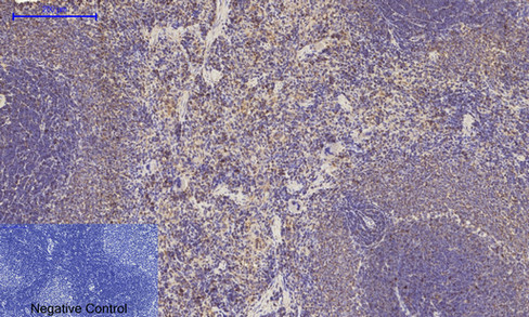
- Immunohistochemical analysis of paraffin-embedded Rat-spleen tissue. 1,JAK1 Polyclonal Antibody was diluted at 1:200(4°C,overnight). 2, Sodium citrate pH 6.0 was used for antibody retrieval(>98°C,20min). 3,Secondary antibody was diluted at 1:200(room tempeRature, 30min). Negative control was used by secondary antibody only.
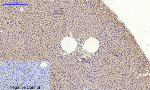
- Immunohistochemical analysis of paraffin-embedded Mouse-liver tissue. 1,JAK1 Polyclonal Antibody was diluted at 1:200(4°C,overnight). 2, Sodium citrate pH 6.0 was used for antibody retrieval(>98°C,20min). 3,Secondary antibody was diluted at 1:200(room tempeRature, 30min). Negative control was used by secondary antibody only.

- Immunohistochemical analysis of paraffin-embedded Mouse-kidney tissue. 1,JAK1 Polyclonal Antibody was diluted at 1:200(4°C,overnight). 2, Sodium citrate pH 6.0 was used for antibody retrieval(>98°C,20min). 3,Secondary antibody was diluted at 1:200(room tempeRature, 30min). Negative control was used by secondary antibody only.
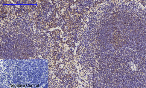
- Immunohistochemical analysis of paraffin-embedded Mouse-spleen tissue. 1,JAK1 Polyclonal Antibody was diluted at 1:200(4°C,overnight). 2, Sodium citrate pH 6.0 was used for antibody retrieval(>98°C,20min). 3,Secondary antibody was diluted at 1:200(room tempeRature, 30min). Negative control was used by secondary antibody only.
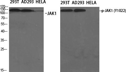
- Western Blot analysis of various cells using JAK1 Polyclonal Antibody diluted at 1:1000
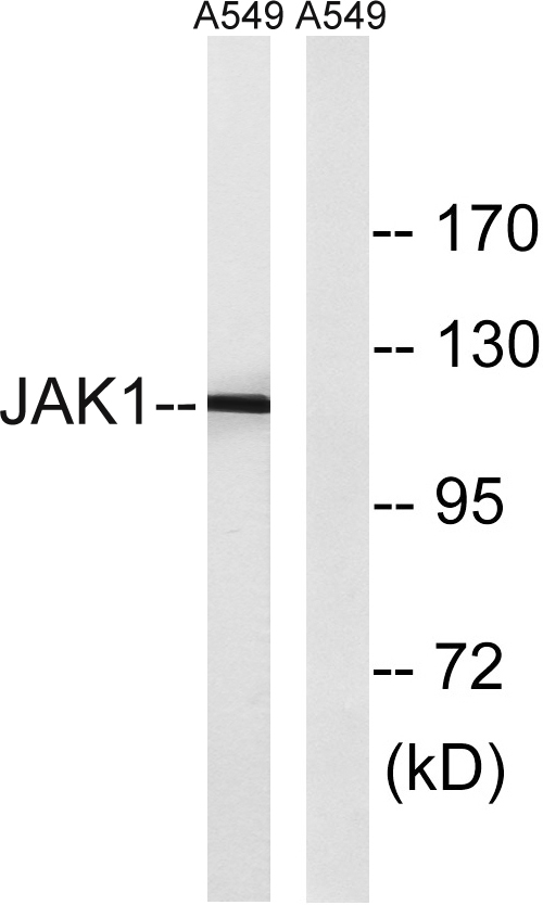
- Western blot analysis of lysates from A549, using JAK1 Antibody. The lane on the right is blocked with the synthesized peptide.


