Histone H1 Polyclonal Antibody
- 货号:YT2150
- 应用:WB;IHC;IF;ELISA
- 种属:Human;Mouse
- 免疫原:
- The antiserum was produced against synthesized peptide derived from human Histone H1. AA range:1-50
- 特异性:
- Histone H1 Polyclonal Antibody detects endogenous levels of Histone H1 protein.
- 组成:
- Liquid in PBS containing 50% glycerol, 0.5% BSA and 0.02% sodium azide.
- 来源:
- Polyclonal, Rabbit,IgG
- 稀释:
- WB 1:500 - 1:2000. IHC 1:100 - 1:300. IF 1:200 - 1:1000. ELISA: 1:20000. Not yet tested in other applications.
- 纯化工艺:
- The antibody was affinity-purified from rabbit antiserum by affinity-chromatography using epitope-specific immunogen.
- 储存:
- -15°C to -25°C/1 year(Do not lower than -25°C)
- 其他名称:
- HIST1H1B;H1F5;Histone H1.5;Histone H1a;Histone H1b;Histone H1s-3;HIST1H1D;H1F3;Histone H1.3;Histone H1c;Histone H1s-2;HIST1H1E;H1F4;Histone H1.4;Histone H1b;Histone H1s-4
- 背景:
- Histones are basic nuclear proteins responsible for nucleosome structure of the chromosomal fiber in eukaryotes. Two molecules of each of the four core histones (H2A, H2B, H3, and H4) form an octamer, around which approximately 146 bp of DNA is wrapped in repeating units, called nucleosomes. The linker histone, H1, interacts with linker DNA between nucleosomes and functions in the compaction of chromatin into higher order structures. This gene is intronless and encodes a replication-dependent histone that is a member of the histone H1 family. Transcripts from this gene lack polyA tails but instead contain a palindromic termination element. This gene is found in the small histone gene cluster on chromosome 6p22-p21.3. [provided by RefSeq, Aug 2015],
- 功能:
- function:Histones H1 are necessary for the condensation of nucleosome chains into higher order structures.,similarity:Belongs to the histone H1/H5 family.,
- 细胞定位:
- Nucleus. Chromosome. According to PubMed:15911621 more commonly found in heterochromatin. According to PubMed:10997781 associates with actively transcribed chromatin and not heterochromatin.
- 组织表达:
- Ubiquitous. Expressed in the majority of the cell lines tested and in testis.
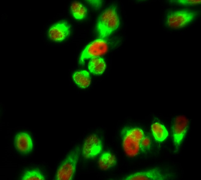
- Immunofluorescence analysis of Hela cell. 1,Histone H1 Polyclonal Antibody(red) was diluted at 1:200(4° overnight). α-SMA Monoclonal Antibody(6A12)(green) was diluted at 1:200(4° overnight). 2, Goat Anti Rabbit Alexa Fluor 594 Catalog:RS3611 was diluted at 1:1000(room temperature, 50min). Goat Anti Mouse Alexa Fluor 488 Catalog:RS3208 was diluted at 1:1000(room temperature, 50min).
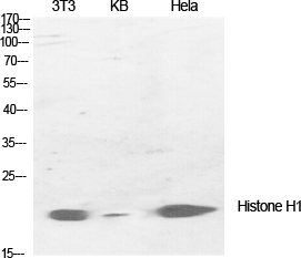
- Western Blot analysis of various cells using Histone H1 Polyclonal Antibody diluted at 1:1000 cells nucleus extracted by Minute TM Cytoplasmic and Nuclear Fractionation kit (SC-003,Inventbiotech,MN,USA).
.jpg)
- Western Blot analysis of 3T3 cells using Histone H1 Polyclonal Antibody diluted at 1:1000 cells nucleus extracted by Minute TM Cytoplasmic and Nuclear Fractionation kit (SC-003,Inventbiotech,MN,USA).
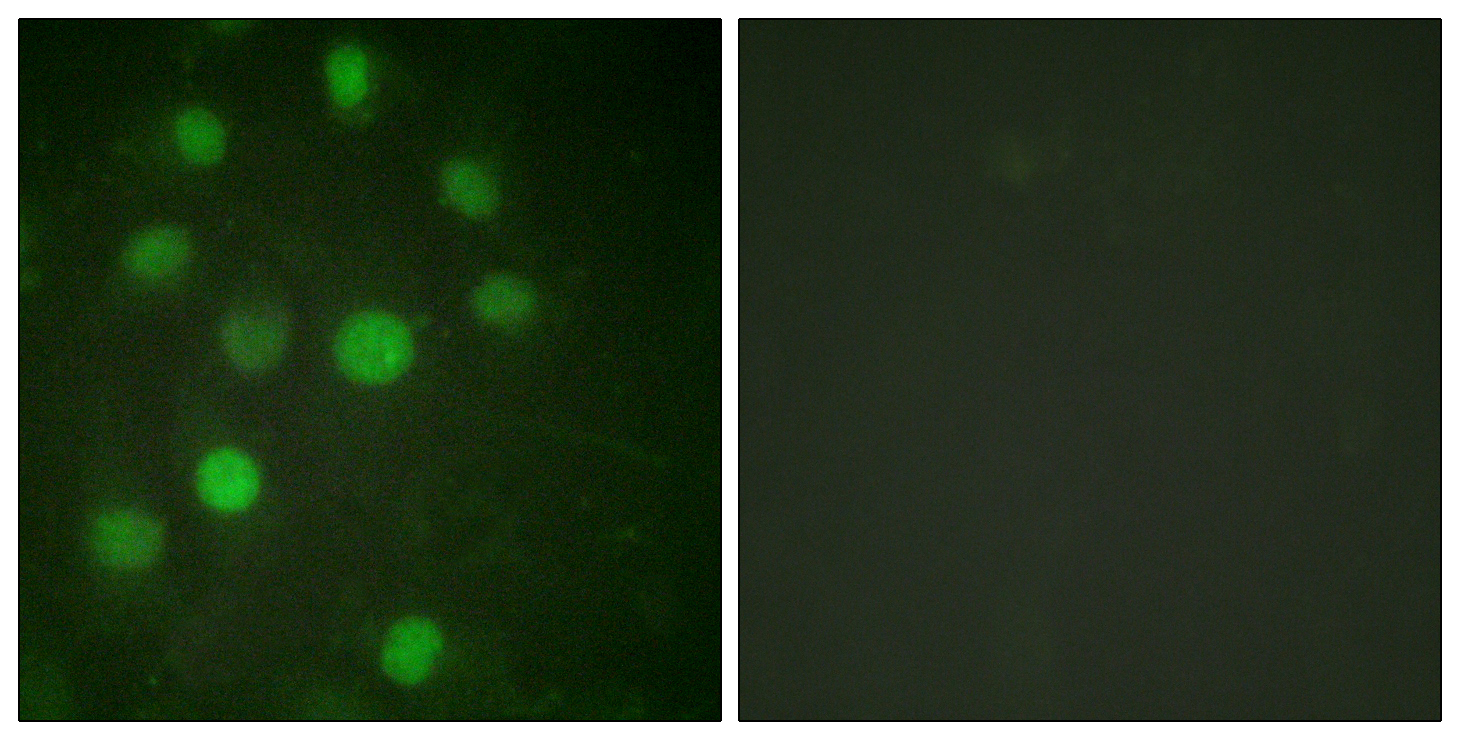
- Immunofluorescence analysis of HUVEC cells, using Histone H1 Antibody. The picture on the right is blocked with the synthesized peptide.
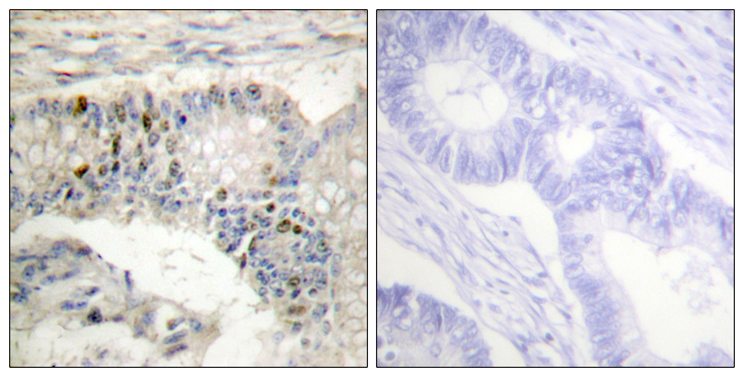
- Immunohistochemistry analysis of paraffin-embedded human colon carcinoma tissue, using Histone H1 Antibody. The picture on the right is blocked with the synthesized peptide.
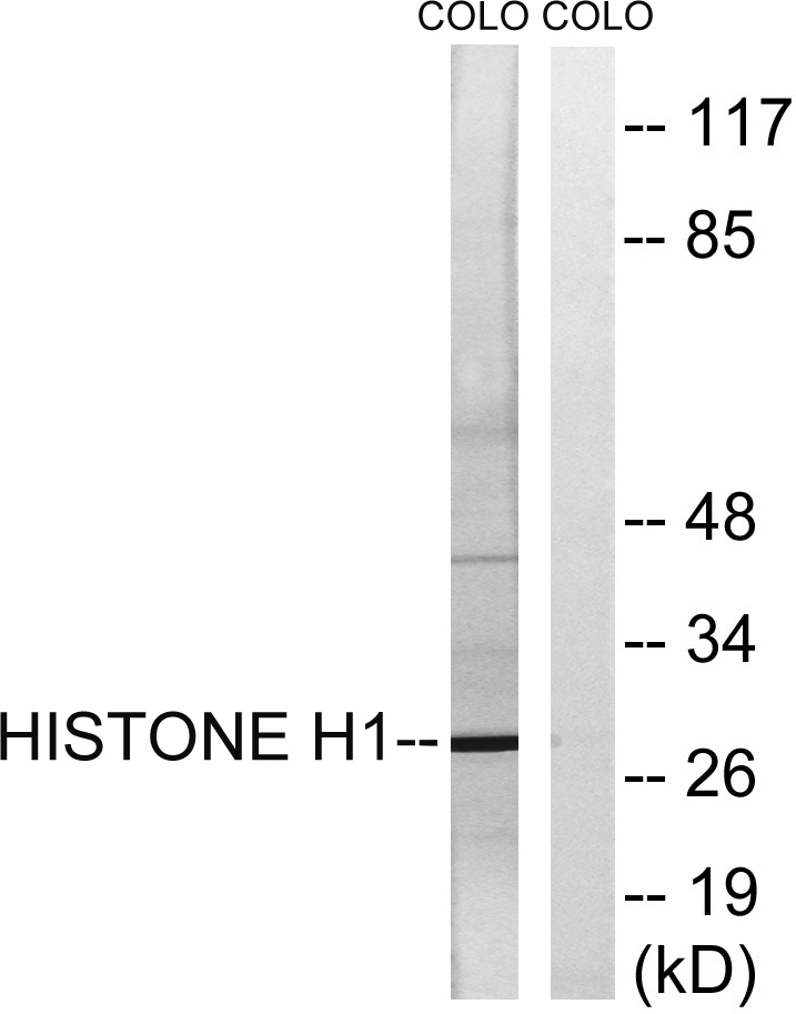
- Western blot analysis of lysates from COLO cells, using Histone H1 Antibody. The lane on the right is blocked with the synthesized peptide.


.jpg)


