- 靶点:
- Estrogen Receptor-β
- 简介:
- >>Endocrine resistance;>>Estrogen signaling pathway;>>Prolactin signaling pathway;>>GnRH secretion;>>Pathways in cancer;>>Chemical carcinogenesis - receptor activation;>>Breast cancer
- 基因名称:
- ESR2
- 蛋白名称:
- Estrogen receptor beta
- Human Gene Id:
- 2100
- Human Swiss Prot No:
- Q92731
- Mouse Gene Id:
- 13983
- Mouse Swiss Prot No:
- O08537
- Rat Gene Id:
- 25149
- Rat Swiss Prot No:
- Q62986
- 免疫原:
- The antiserum was produced against synthesized peptide derived from human Estrogen Receptor-beta. AA range:71-120
- 特异性:
- ERβ Polyclonal Antibody detects endogenous levels of ERβ protein.
- 组成:
- Liquid in PBS containing 50% glycerol, 0.5% BSA and 0.02% sodium azide.
- 来源:
- Polyclonal, Rabbit,IgG
- 稀释:
- WB 1:500-2000, IF 1:50-300, IHC 1:50-300
- 纯化工艺:
- The antibody was affinity-purified from rabbit antiserum by affinity-chromatography using epitope-specific immunogen.
- 浓度:
- 1 mg/ml
- 储存:
- -15°C to -25°C/1 year(Do not lower than -25°C)
- 其他名称:
- ESR2;ESTRB;NR3A2;Estrogen receptor beta;ER-beta;Nuclear receptor subfamily 3 group A member 2
- 实测条带:
- 59kD
- 背景:
- This gene encodes a member of the family of estrogen receptors and superfamily of nuclear receptor transcription factors. The gene product contains an N-terminal DNA binding domain and C-terminal ligand binding domain and is localized to the nucleus, cytoplasm, and mitochondria. Upon binding to 17beta-estradiol or related ligands, the encoded protein forms homo- or hetero-dimers that interact with specific DNA sequences to activate transcription. Some isoforms dominantly inhibit the activity of other estrogen receptor family members. Several alternatively spliced transcript variants of this gene have been described, but the full-length nature of some of these variants has not been fully characterized. [provided by RefSeq, Jul 2008],
- 功能:
- domain:Composed of three domains: a modulating N-terminal domain, a DNA-binding domain and a C-terminal steroid-binding domain.,function:Nuclear hormone receptor. Binds estrogens with an affinity similar to that of ESR1, and activates expression of reporter genes containing estrogen response elements (ERE) in an estrogen-dependent manner. Isoform beta-cx lacks ligand binding ability and has no or only very low ere binding activity resulting in the loss of ligand-dependent transactivation ability. DNA-binding by ESR1 and ESR2 is rapidly lost at 37 degrees Celsius in the absence of ligand while in the presence of 17 beta-estradiol and 4-hydroxy-tamoxifen loss in DNA-binding at elevated temperature is more gradual.,online information:Estrogen receptor entry,similarity:Belongs to the nuclear hormone receptor family.,similarity:Belongs to the nuclear hormone receptor family. NR3 subfamily.,si
- 细胞定位:
- Nucleus .
- 组织表达:
- [Isoform 1]: Expressed in testis and ovary, and at a lower level in heart, brain, placenta, liver, skeletal muscle, spleen, thymus, prostate, colon, bone marrow, mammary gland and uterus. Also found in uterine bone, breast, and ovarian tumor cell lines, but not in colon and liver tumors. ; [Isoform 2]: Expressed in spleen, thymus, testis and ovary and at a lower level in skeletal muscle, prostate, colon, small intestine, leukocytes, bone marrow, mammary gland and uterus. ; [Isoform 4]: Expressed in the testis. ; [Isoform 5]: Expressed in testis, and at a lower level in spleen, thymus, ovary, mammary gland and uterus. ; [Isoform 6]: Expressed in testis, placenta, skeletal muscle, spleen and leukocytes, and at a lower level in heart, lung, liver, kidney, pancreas, thymus, prostate, colon, sm
Proteomic analysis reveals that cigarette smoke exposure diminishes ovarian reserve in mice by disrupting the CREB1-mediated ovarian granulosa cell proliferation-apoptosis balance ECOTOXICOLOGY AND ENVIRONMENTAL SAFETY Mengting Xu WB Mouse,Human 1:2000 ovarian tissue KGN cell
货号:YT1637
- June 19-2018
- WESTERN IMMUNOBLOTTING PROTOCOL
- June 19-2018
- IMMUNOHISTOCHEMISTRY-PARAFFIN PROTOCOL
- June 19-2018
- IMMUNOFLUORESCENCE PROTOCOL
- September 08-2020
- FLOW-CYTOMEYRT-PROTOCOL
- May 20-2022
- Cell-Based ELISA│解您多样本WB检测之困扰
- July 13-2018
- CELL-BASED-ELISA-PROTOCOL-FOR-ACETYL-PROTEIN
- July 13-2018
- CELL-BASED-ELISA-PROTOCOL-FOR-PHOSPHO-PROTEIN
- July 13-2018
- Antibody-FAQs
- 产品图片
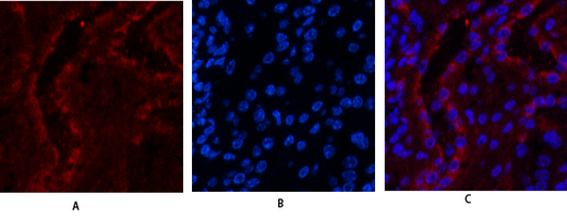
- Immunofluorescence analysis of mouse-kidney tissue. 1,ERβ Polyclonal Antibody(red) was diluted at 1:200(4° overnight). 2, Cy3 labled Secondary antibody was diluted at 1:300(room temperature, 50min).3, Picture B: DAPI(blue) 10min. Picture A:Target. Picture B: DAPI. Picture C: merge of A+B
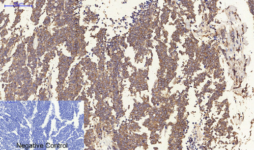
- Immunohistochemical analysis of paraffin-embedded Human-lung-cancer tissue. 1,ERβ Polyclonal Antibody was diluted at 1:200(4°C,overnight). 2, Sodium citrate pH 6.0 was used for antibody retrieval(>98°C,20min). 3,Secondary antibody was diluted at 1:200(room tempeRature, 30min). Negative control was used by secondary antibody only.
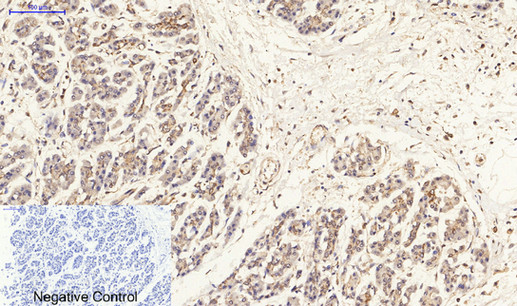
- Immunohistochemical analysis of paraffin-embedded Human-stomach-cancer tissue. 1,ERβ Polyclonal Antibody was diluted at 1:200(4°C,overnight). 2, Sodium citrate pH 6.0 was used for antibody retrieval(>98°C,20min). 3,Secondary antibody was diluted at 1:200(room tempeRature, 30min). Negative control was used by secondary antibody only.
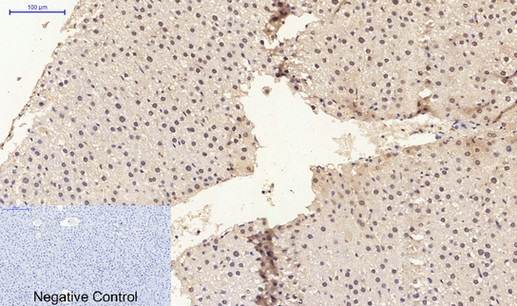
- Immunohistochemical analysis of paraffin-embedded Rat-liver tissue. 1,ERβ Polyclonal Antibody was diluted at 1:200(4°C,overnight). 2, Sodium citrate pH 6.0 was used for antibody retrieval(>98°C,20min). 3,Secondary antibody was diluted at 1:200(room tempeRature, 30min). Negative control was used by secondary antibody only.
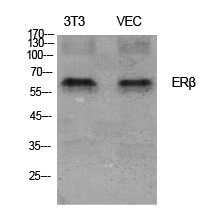
- Western Blot analysis of various cells using ERβ Polyclonal Antibody diluted at 1:1000
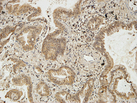
- Immunohistochemical analysis of paraffin-embedded Human lung cancer. 1, Antibody was diluted at 1:100(4° overnight). 2, High-pressure and temperature EDTA, pH8.0 was used for antigen retrieval. 3,Secondary antibody was diluted at 1:200(room temperature, 30min).
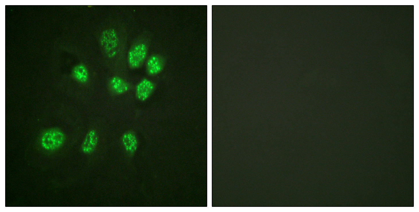
- Immunofluorescence analysis of HeLa cells, using Estrogen Receptor-beta Antibody. The picture on the right is blocked with the synthesized peptide.
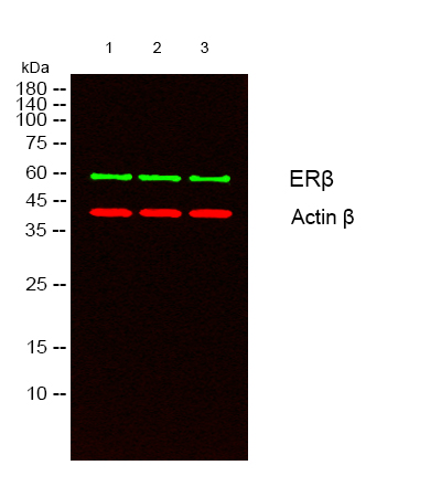
- Western blot analysis of lysates from 1)3T3 , 2) VEC , 3) HeLa cells, (Green) primary antibody was diluted at 1:1000, 4°over night, secondary antibody(cat:RS23920)was diluted at 1:10000, 37° 1hour. (Red) Actin β Monoclonal Antibody(5B7) (cat:YM3028) antibody was diluted at 1:5000 as loading control, 4° over night,secondary antibody(cat:RS23710)was diluted at 1:10000, 37° 1hour.


