- 靶点:
- c-Src
- 简介:
- >>EGFR tyrosine kinase inhibitor resistance;>>Endocrine resistance;>>ErbB signaling pathway;>>Rap1 signaling pathway;>>Chemokine signaling pathway;>>Mitophagy - animal;>>Endocytosis;>>Axon guidance;>>VEGF signaling pathway;>>Focal adhesion;>>Adherens junction;>>Tight junction;>>Gap junction;>>Platelet activation;>>Neutrophil extracellular trap formation;>>C-type lectin receptor signaling pathway;>>GABAergic synapse;>>Inflammatory mediator regulation of TRP channels;>>Regulation of actin cytoskeleton;>>GnRH signaling pathway;>>Estrogen signaling pathway;>>Prolactin signaling pathway;>>Thyroid hormone signaling pathway;>>Oxytocin signaling pathway;>>Relaxin signaling pathway;>>Bacterial invasion of epithelial cells;>>Epithelial cell signaling in Helicobacter pylori infection;>>Pathogenic Escherichia coli infection;>>Shigellosis;>>Yersinia infection;>>Tuberculosis;>>Hepatitis B;>>Human cytomegalovirus infection;>>Kaposi sarcoma-associated herpesvirus infection;>>Herpes simplex virus 1 inf
- 基因名称:
- SRC
- 蛋白名称:
- Proto-oncogene tyrosine-protein kinase Src
- Human Gene Id:
- 6714
- Human Swiss Prot No:
- P12931
- Mouse Gene Id:
- 20779
- Mouse Swiss Prot No:
- P05480
- Rat Swiss Prot No:
- Q9WUD9
- 免疫原:
- Synthesized peptide derived from c-Src . at AA range: 360-440
- 特异性:
- c-Src Polyclonal Antibody detects endogenous levels of c-Src protein.
- 组成:
- Liquid in PBS containing 50% glycerol, 0.5% BSA and 0.02% sodium azide.
- 来源:
- Polyclonal, Rabbit,IgG
- 稀释:
- WB 1:500 - 1:2000. IHC 1:100 - 1:300. IF 1:200 - 1:1000. ELISA: 1:40000. Not yet tested in other applications.
- 纯化工艺:
- The antibody was affinity-purified from rabbit antiserum by affinity-chromatography using epitope-specific immunogen.
- 浓度:
- 1 mg/ml
- 储存:
- -15°C to -25°C/1 year(Do not lower than -25°C)
- 其他名称:
- SRC;SRC1;Proto-oncogene tyrosine-protein kinase Src;Proto-oncogene c-Src;pp60c-src;p60-Src
- 实测条带:
- 60kD
- 背景:
- This gene is highly similar to the v-src gene of Rous sarcoma virus. This proto-oncogene may play a role in the regulation of embryonic development and cell growth. The protein encoded by this gene is a tyrosine-protein kinase whose activity can be inhibited by phosphorylation by c-SRC kinase. Mutations in this gene could be involved in the malignant progression of colon cancer. Two transcript variants encoding the same protein have been found for this gene. [provided by RefSeq, Jul 2008],
- 功能:
- catalytic activity:ATP + a [protein]-L-tyrosine = ADP + a [protein]-L-tyrosine phosphate.,PTM:Phosphorylated on Tyr-530 by c-Src kinase (CSK). The phosphorylated form is termed pp60c-src. The phosphorylated tail interacts with the SH2 domain thereby repressing kinase activity.,similarity:Belongs to the protein kinase superfamily. Tyr protein kinase family. SRC subfamily.,similarity:Contains 1 protein kinase domain.,similarity:Contains 1 SH2 domain.,similarity:Contains 1 SH3 domain.,subunit:Interacts with DDEF1/ASAP1; via the SH3 domain. Interacts with CCPG1 (By similarity). Interacts with CDCP1, PELP1, TGFB1I1 and TOM1L2. Interacts with the cytoplasmic domain of MUC1, phosphorylates it and increases binding of MUC1 with beta-catenin. Interacts with RALGPS1; via the SH3 domain. Interacts with HEV ORF3 protein; via the SH3 domain.,
- 细胞定位:
- Cell membrane ; Lipid-anchor . Mitochondrion inner membrane . Nucleus . Cytoplasm, cytoskeleton . Cytoplasm, perinuclear region . Cell junction, focal adhesion . Localizes to focal adhesion sites following integrin engagement (PubMed:22801373). Localization to focal adhesion sites requires myristoylation and the SH3 domain (PubMed:7525268). Colocalizes with PDLIM4 at the perinuclear region, but not at focal adhesions (PubMed:19307596). .
- 组织表达:
- Expressed ubiquitously. Platelets, neurons and osteoclasts express 5-fold to 200-fold higher levels than most other tissues.; [Isoform 1]: Expressed in spleen and liver. ; [Isoform 2]: Expressed in brain. ; [Isoform 3]: Expressed in brain.
CX3CL1 promotes tumour cell by inducing tyrosine phosphorylation of cortactin in lung cancer. JOURNAL OF CELLULAR AND MOLECULAR MEDICINE J Cell Mol Med. 2021 Jan;25(1):132-146 WB Human 1:1000 A549 cell, H520 cell
货号:YT1136
Lu, Qian, et al. "Norisoboldine suppresses VEGF-induced endothelial cell migration via the cAMP-PKA-NF-κB/Notch1 pathway." PloS one 8.12 (2013): e81220.
货号:YT1136
Lu, Jian, et al. "Role of LKB1 in migration and invasion of Cr (VI)-transformed human bronchial epithelial Beas-2B cells." Anti-cancer drugs 29.7 (2018): 660-673.
货号:YT1136
Gut microbial co-metabolite 2-methylbutyrylcarnitine exacerbates thrombosis via binding to and activating integrin α2β1 Cell Metabolism Kan Huang WB Rat 1:1000 plasma
货号:YT1136
PDGFRα+ITGA11+ fibroblasts foster early-stage cancer lymphovascular invasion and lymphatic metastasis via ITGA11-SELE interplay CANCER CELL Hanhao Zheng CoIP Human plasma human LECs (HLECs)
货号:YT1136
- June 19-2018
- WESTERN IMMUNOBLOTTING PROTOCOL
- June 19-2018
- IMMUNOHISTOCHEMISTRY-PARAFFIN PROTOCOL
- June 19-2018
- IMMUNOFLUORESCENCE PROTOCOL
- September 08-2020
- FLOW-CYTOMEYRT-PROTOCOL
- May 20-2022
- Cell-Based ELISA│解您多样本WB检测之困扰
- July 13-2018
- CELL-BASED-ELISA-PROTOCOL-FOR-ACETYL-PROTEIN
- July 13-2018
- CELL-BASED-ELISA-PROTOCOL-FOR-PHOSPHO-PROTEIN
- July 13-2018
- Antibody-FAQs
- 产品图片

- Immunofluorescence analysis of rat-spleen tissue. 1,c-Src Polyclonal Antibody(red) was diluted at 1:200(4°C,overnight). 2, Cy3 labled Secondary antibody was diluted at 1:300(room temperature, 50min).3, Picture B: DAPI(blue) 10min. Picture A:Target. Picture B: DAPI. Picture C: merge of A+B
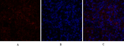
- Immunofluorescence analysis of rat-spleen tissue. 1,c-Src Polyclonal Antibody(red) was diluted at 1:200(4°C,overnight). 2, Cy3 labled Secondary antibody was diluted at 1:300(room temperature, 50min).3, Picture B: DAPI(blue) 10min. Picture A:Target. Picture B: DAPI. Picture C: merge of A+B
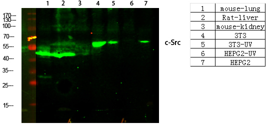
- Western Blot analysis of various cells using primary antibody diluted at 1:1000(4°C overnight). Secondary antibody:Goat Anti-rabbit IgG IRDye 800( diluted at 1:5000, 25°C, 1 hour)
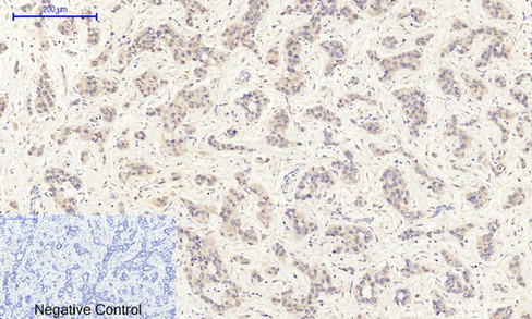
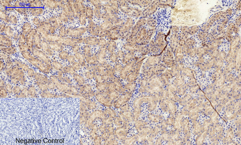
- Immunohistochemical analysis of paraffin-embedded Human-liver-cancer tissue. 1,c-Src Polyclonal Antibody was diluted at 1:200(4°C,overnight). 2, Sodium citrate pH 6.0 was used for antibody retrieval(>98°C,20min). 3,Secondary antibody was diluted at 1:200(room tempeRature, 30min). Negative control was used by secondary antibody only.

- Immunohistochemical analysis of paraffin-embedded Human-stomach tissue. 1,c-Src Polyclonal Antibody was diluted at 1:200(4°C,overnight). 2, Sodium citrate pH 6.0 was used for antibody retrieval(>98°C,20min). 3,Secondary antibody was diluted at 1:200(room tempeRature, 30min). Negative control was used by secondary antibody only.
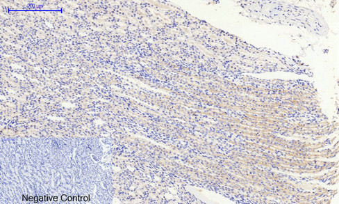
- Immunohistochemical analysis of paraffin-embedded Rat-kidney tissue. 1,c-Src Polyclonal Antibody was diluted at 1:200(4°C,overnight). 2, Sodium citrate pH 6.0 was used for antibody retrieval(>98°C,20min). 3,Secondary antibody was diluted at 1:200(room tempeRature, 30min). Negative control was used by secondary antibody only.
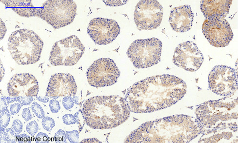
- Immunohistochemical analysis of paraffin-embedded Mouse-testis tissue. 1,c-Src Polyclonal Antibody was diluted at 1:200(4°C,overnight). 2, Sodium citrate pH 6.0 was used for antibody retrieval(>98°C,20min). 3,Secondary antibody was diluted at 1:200(room tempeRature, 30min). Negative control was used by secondary antibody only.
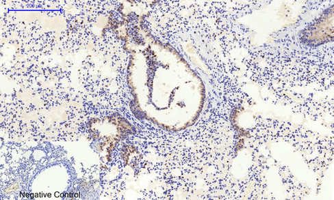
- Immunohistochemical analysis of paraffin-embedded Mouse-lung tissue. 1,c-Src Polyclonal Antibody was diluted at 1:200(4°C,overnight). 2, Sodium citrate pH 6.0 was used for antibody retrieval(>98°C,20min). 3,Secondary antibody was diluted at 1:200(room tempeRature, 30min). Negative control was used by secondary antibody only.
- Immunohistochemical analysis of paraffin-embedded Mouse-kidney tissue. 1,c-Src Polyclonal Antibody was diluted at 1:200(4°C,overnight). 2, Sodium citrate pH 6.0 was used for antibody retrieval(>98°C,20min). 3,Secondary antibody was diluted at 1:200(room tempeRature, 30min). Negative control was used by secondary antibody only.
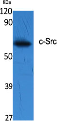
- Western Blot analysis of various cells using c-Src Polyclonal Antibody diluted at 1:2000
.jpg)
- Western Blot analysis of 293 cells using c-Src Polyclonal Antibody diluted at 1:2000


