- 靶点:
- Claudin-5
- 简介:
- >>Cell adhesion molecules;>>Tight junction;>>Leukocyte transendothelial migration;>>Pathogenic Escherichia coli infection;>>Hepatitis C
- 基因名称:
- CLDN5
- 蛋白名称:
- Claudin-5
- Human Gene Id:
- 7122
- Human Swiss Prot No:
- O00501
- Mouse Gene Id:
- 12741
- Mouse Swiss Prot No:
- O54942
- Rat Gene Id:
- 65131
- Rat Swiss Prot No:
- Q9JKD6
- 免疫原:
- The antiserum was produced against synthesized peptide derived from human Claudin 5. AA range:169-218
- 特异性:
- Claudin-5 Polyclonal Antibody detects endogenous levels of Claudin-5 protein.
- 组成:
- Liquid in PBS containing 50% glycerol, 0.5% BSA and 0.02% sodium azide.
- 来源:
- Polyclonal, Rabbit,IgG
- 稀释:
- WB 1:500 - 1:2000. IHC: 1:100-300 ELISA: 1:20000. IF 1:100-300 Not yet tested in other applications.
- 纯化工艺:
- The antibody was affinity-purified from rabbit antiserum by affinity-chromatography using epitope-specific immunogen.
- 浓度:
- 1 mg/ml
- 储存:
- -15°C to -25°C/1 year(Do not lower than -25°C)
- 其他名称:
- CLDN5;AWAL;TMVCF;Claudin-5;Transmembrane protein deleted in VCFS;TMDVCF
- 实测条带:
- 23kD
- 背景:
- This gene encodes a member of the claudin family. Claudins are integral membrane proteins and components of tight junction strands. Tight junction strands serve as a physical barrier to prevent solutes and water from passing freely through the paracellular space between epithelial or endothelial cell sheets. Mutations in this gene have been found in patients with velocardiofacial syndrome. Alternatively spliced transcript variants encoding the same protein have been found for this gene. [provided by RefSeq, Aug 2008],
- 功能:
- function:Plays a major role in tight junction-specific obliteration of the intercellular space.,similarity:Belongs to the claudin family.,subunit:Directly interacts with TJP1/ZO-1, TJP2/ZO-2 and TJP3/ZO-3. Interacts with MPDZ.,
- 细胞定位:
- Cell junction, tight junction. Cell membrane; Multi-pass membrane protein.
- 组织表达:
- Adipose tissue,Brain,Lung,Prostate,
Quercetin attenuates ischemia reperfusion injury by protecting the blood-brain barrier through Sirt1 in MCAO rats. JOURNAL OF ASIAN NATURAL PRODUCTS RESEARCH J Asian Nat Prod Res. 2022;24(3):278-289 WB Rat Brain
货号:YT0953
VEGF-A in serum protects against memory impairment in APP/PS1 transgenic mice by blocking neutrophil infiltration. Kaihua Guo IF Mouse 1:400 hippocampus
货号:YT0953
- June 19-2018
- WESTERN IMMUNOBLOTTING PROTOCOL
- June 19-2018
- IMMUNOHISTOCHEMISTRY-PARAFFIN PROTOCOL
- June 19-2018
- IMMUNOFLUORESCENCE PROTOCOL
- September 08-2020
- FLOW-CYTOMEYRT-PROTOCOL
- May 20-2022
- Cell-Based ELISA│解您多样本WB检测之困扰
- July 13-2018
- CELL-BASED-ELISA-PROTOCOL-FOR-ACETYL-PROTEIN
- July 13-2018
- CELL-BASED-ELISA-PROTOCOL-FOR-PHOSPHO-PROTEIN
- July 13-2018
- Antibody-FAQs
- 产品图片
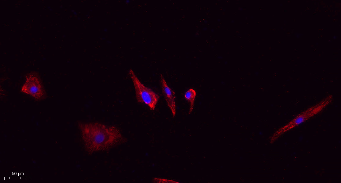
- Immunofluorescence analysis of A549. 1,primary Antibody(red) was diluted at 1:200(4°C overnight). 2, Goat Anti Rabbit IgG (H&L) - Alexa Fluor 594 Secondary antibody was diluted at 1:1000(room temperature, 50min).3, Picture B: DAPI(blue) 10min.
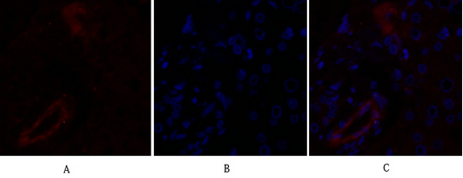
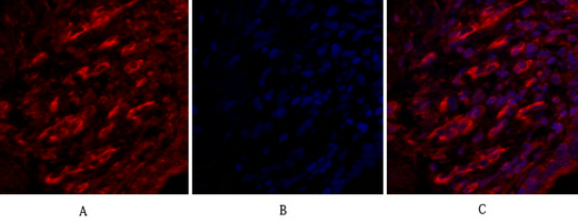
- Immunofluorescence analysis of human-liver tissue. 1,Claudin-5 Polyclonal Antibody(red) was diluted at 1:200(4°C,overnight). 2, Cy3 labled Secondary antibody was diluted at 1:300(room temperature, 50min).3, Picture B: DAPI(blue) 10min. Picture A:Target. Picture B: DAPI. Picture C: merge of A+B
- Immunofluorescence analysis of human-liver tissue. 1,Claudin-5 Polyclonal Antibody(red) was diluted at 1:200(4°C,overnight). 2, Cy3 labled Secondary antibody was diluted at 1:300(room temperature, 50min).3, Picture B: DAPI(blue) 10min. Picture A:Target. Picture B: DAPI. Picture C: merge of A+B
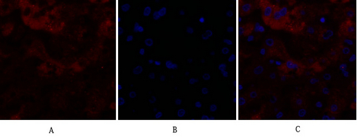
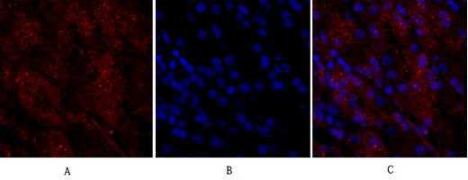
- Immunofluorescence analysis of human-lung tissue. 1,Claudin-5 Polyclonal Antibody(red) was diluted at 1:200(4°C,overnight). 2, Cy3 labled Secondary antibody was diluted at 1:300(room temperature, 50min).3, Picture B: DAPI(blue) 10min. Picture A:Target. Picture B: DAPI. Picture C: merge of A+B
- Immunofluorescence analysis of human-lung tissue. 1,Claudin-5 Polyclonal Antibody(red) was diluted at 1:200(4°C,overnight). 2, Cy3 labled Secondary antibody was diluted at 1:300(room temperature, 50min).3, Picture B: DAPI(blue) 10min. Picture A:Target. Picture B: DAPI. Picture C: merge of A+B
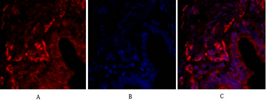
- Immunofluorescence analysis of human-stomach tissue. 1,Claudin-5 Polyclonal Antibody(red) was diluted at 1:200(4°C,overnight). 2, Cy3 labled Secondary antibody was diluted at 1:300(room temperature, 50min).3, Picture B: DAPI(blue) 10min. Picture A:Target. Picture B: DAPI. Picture C: merge of A+B
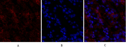
- Immunofluorescence analysis of human-stomach tissue. 1,Claudin-5 Polyclonal Antibody(red) was diluted at 1:200(4°C,overnight). 2, Cy3 labled Secondary antibody was diluted at 1:300(room temperature, 50min).3, Picture B: DAPI(blue) 10min. Picture A:Target. Picture B: DAPI. Picture C: merge of A+B
- Immunohistochemical analysis of paraffin-embedded Human-uterus tissue. 1,Claudin-5 Polyclonal Antibody was diluted at 1:200(4°C,overnight). 2, Sodium citrate pH 6.0 was used for antibody retrieval(>98°C,20min). 3,Secondary antibody was diluted at 1:200(room tempeRature, 30min). Negative control was used by secondary antibody only.
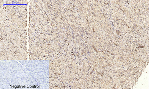
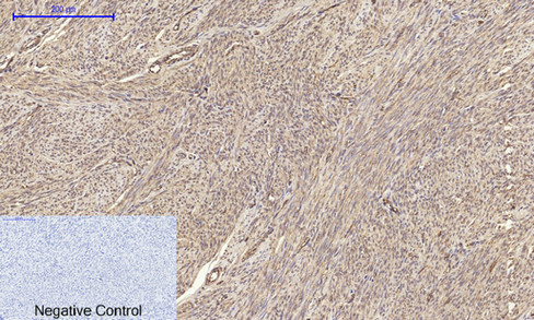
- Immunohistochemical analysis of paraffin-embedded Human-uterus-cancer tissue. 1,Claudin-5 Polyclonal Antibody was diluted at 1:200(4°C,overnight). 2, Sodium citrate pH 6.0 was used for antibody retrieval(>98°C,20min). 3,Secondary antibody was diluted at 1:200(room tempeRature, 30min). Negative control was used by secondary antibody only.
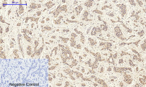
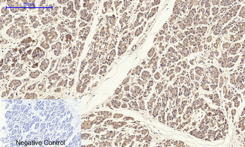
- Immunohistochemical analysis of paraffin-embedded Human-liver-cancer tissue. 1,Claudin-5 Polyclonal Antibody was diluted at 1:200(4°C,overnight). 2, Sodium citrate pH 6.0 was used for antibody retrieval(>98°C,20min). 3,Secondary antibody was diluted at 1:200(room tempeRature, 30min). Negative control was used by secondary antibody only.
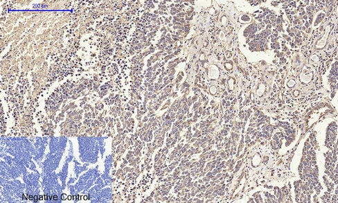
- Immunohistochemical analysis of paraffin-embedded Human-lung-cancer tissue. 1,Claudin-5 Polyclonal Antibody was diluted at 1:200(4°C,overnight). 2, Sodium citrate pH 6.0 was used for antibody retrieval(>98°C,20min). 3,Secondary antibody was diluted at 1:200(room tempeRature, 30min). Negative control was used by secondary antibody only.
- Immunohistochemical analysis of paraffin-embedded Human-stomach tissue. 1,Claudin-5 Polyclonal Antibody was diluted at 1:200(4°C,overnight). 2, Sodium citrate pH 6.0 was used for antibody retrieval(>98°C,20min). 3,Secondary antibody was diluted at 1:200(room tempeRature, 30min). Negative control was used by secondary antibody only.
- Immunohistochemical analysis of paraffin-embedded Human-stomach-cancer tissue. 1,Claudin-5 Polyclonal Antibody was diluted at 1:200(4°C,overnight). 2, Sodium citrate pH 6.0 was used for antibody retrieval(>98°C,20min). 3,Secondary antibody was diluted at 1:200(room tempeRature, 30min). Negative control was used by secondary antibody only.
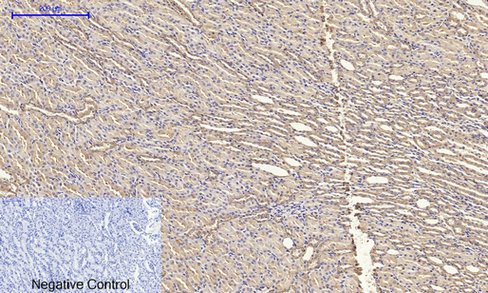
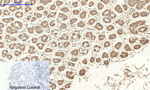
- Immunohistochemical analysis of paraffin-embedded Rat-kidney tissue. 1,Claudin-5 Polyclonal Antibody was diluted at 1:200(4°C,overnight). 2, Sodium citrate pH 6.0 was used for antibody retrieval(>98°C,20min). 3,Secondary antibody was diluted at 1:200(room tempeRature, 30min). Negative control was used by secondary antibody only.
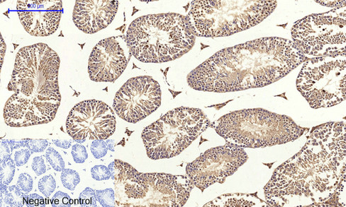
- Immunohistochemical analysis of paraffin-embedded Mouse-testis tissue. 1,Claudin-5 Polyclonal Antibody was diluted at 1:200(4°C,overnight). 2, Sodium citrate pH 6.0 was used for antibody retrieval(>98°C,20min). 3,Secondary antibody was diluted at 1:200(room tempeRature, 30min). Negative control was used by secondary antibody only.
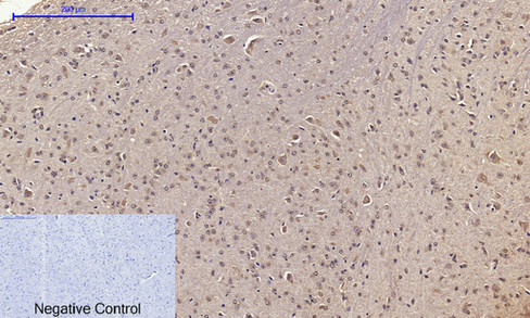
- Immunohistochemical analysis of paraffin-embedded Mouse-brain tissue. 1,Claudin-5 Polyclonal Antibody was diluted at 1:200(4°C,overnight). 2, Sodium citrate pH 6.0 was used for antibody retrieval(>98°C,20min). 3,Secondary antibody was diluted at 1:200(room tempeRature, 30min). Negative control was used by secondary antibody only.
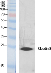
- Western Blot analysis of various cells using Claudin-5 Polyclonal Antibody diluted at 1:500
.jpg)
- Western Blot analysis of A549 cells using Claudin-5 Polyclonal Antibody diluted at 1:500
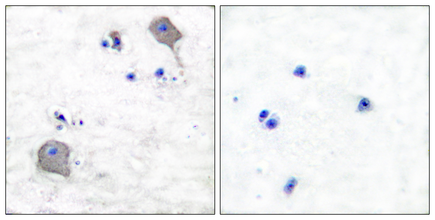
- Immunohistochemistry analysis of paraffin-embedded human brain tissue, using Claudin 5 Antibody. The picture on the right is blocked with the synthesized peptide.
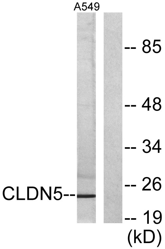
- Western blot analysis of lysates from A549 cells, using Claudin 5 Antibody. The lane on the right is blocked with the synthesized peptide.


