CIP29 Polyclonal Antibody
- 货号:YT0930
- 应用:WB;IHC;IF;ELISA
- 种属:Human;Mouse;Rat
- 蛋白名称:
- SAP domain-containing ribonucleoprotein
- 免疫原:
- The antiserum was produced against synthesized peptide derived from human HCC1. AA range:147-196
- 特异性:
- CIP29 Polyclonal Antibody detects endogenous levels of CIP29 protein.
- 组成:
- Liquid in PBS containing 50% glycerol, 0.5% BSA and 0.02% sodium azide.
- 来源:
- Polyclonal, Rabbit,IgG
- 稀释:
- WB 1:500 - 1:2000. IHC 1:100 - 1:300. ELISA: 1:10000.. IF 1:50-200
- 纯化工艺:
- The antibody was affinity-purified from rabbit antiserum by affinity-chromatography using epitope-specific immunogen.
- 储存:
- -15°C to -25°C/1 year(Do not lower than -25°C)
- 其他名称:
- SARNP;HCC1;HSPC316;SAP domain-containing ribonucleoprotein;Cytokine-induced protein of 29 kDa;Nuclear protein Hcc-1;Proliferation-associated cytokine-inducible protein CIP29
- 背景:
- This gene encodes a protein that is upregulated in response to various cytokines. The encoded protein may play a role in cell cycle progression. A translocation between this gene and the myeloid/lymphoid leukemia gene, resulting in expression of a chimeric protein, has been associated with acute myelomonocytic leukemia. Pseudogenes exist on chromosomes 7 and 8. Alternatively spliced transcript variants have been described. [provided by RefSeq, Feb 2009],
- 功能:
- transcription, regulation of transcription, DNA-dependent, regulation of translation, posttranscriptional regulation of gene expression, regulation of cellular protein metabolic process, regulation of transcription, regulation of RNA metabolic process,
- 细胞定位:
- Nucleus. Nucleus speckle.
- 组织表达:
- Low expression in spleen, liver, pancreas, testis, thymus, heart, and kidney. Increased levels are seen in hepatocellular carcinoma and pancreatic adenocarcinoma.
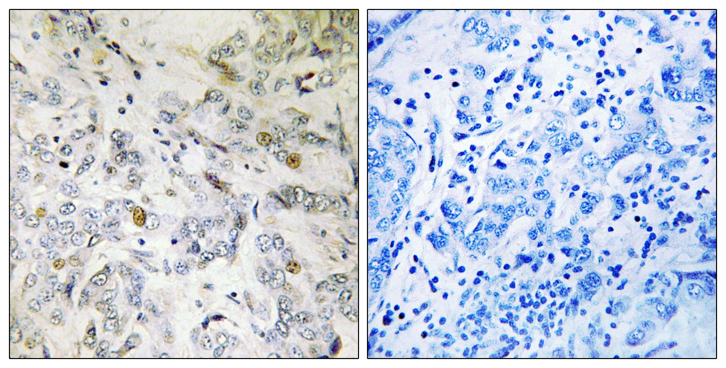
- Immunohistochemistry analysis of paraffin-embedded human breast carcinoma tissue, using HCC1 Antibody. The picture on the right is blocked with the synthesized peptide.
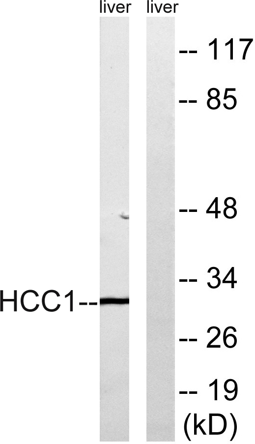
- Western blot analysis of lysates from mouse liver, using HCC1 Antibody. The lane on the right is blocked with the synthesized peptide.


