TAL1 Polyclonal Antibody
- Catalog No.:YT4538
- Applications:WB;IHC;IF;ELISA
- Reactivity:Human;Mouse
- Target:
- TAL1
- Gene Name:
- TAL1
- Protein Name:
- T-cell acute lymphocytic leukemia protein 1
- Human Gene Id:
- 6886
- Human Swiss Prot No:
- P17542
- Mouse Gene Id:
- 21349
- Mouse Swiss Prot No:
- P22091
- Immunogen:
- The antiserum was produced against synthesized peptide derived from human TAL-1. AA range:96-145
- Specificity:
- TAL1 Polyclonal Antibody detects endogenous levels of TAL1 protein.
- Formulation:
- Liquid in PBS containing 50% glycerol, 0.5% BSA and 0.02% sodium azide.
- Source:
- Polyclonal, Rabbit,IgG
- Dilution:
- WB 1:500 - 1:2000. IHC 1:100 - 1:300. ELISA: 1:20000.. IF 1:50-200
- Purification:
- The antibody was affinity-purified from rabbit antiserum by affinity-chromatography using epitope-specific immunogen.
- Concentration:
- 1 mg/ml
- Storage Stability:
- -15°C to -25°C/1 year(Do not lower than -25°C)
- Other Name:
- TAL1;BHLHA17;SCL;TCL5;T-cell acute lymphocytic leukemia protein 1;TAL-1;Class A basic helix-loop-helix protein 17;bHLHa17;Stem cell protein;T-cell leukemia/lymphoma protein 5
- Observed Band(KD):
- 45kD
- Background:
- alternative products:The splicing pattern is cell-lineage dependent,disease:A chromosomal aberration involving TAL1 may be a cause of some T-cell acute lymphoblastic leukemias (T-ALL). Translocation t(1;14)(p32;q11) with T-cell receptor alpha chain (TCRA) genes.,domain:The helix-loop-helix domain is necessary and sufficient for the interaction with DRG1.,function:Implicated in the genesis of hemopoietic malignancies. It may play an important role in hemopoietic differentiation. Serves as a positive regulator of erythroid differentiation.,PTM:Phosphorylated on serine residues. Phosphorylation of Ser-122 is strongly stimulated by hypoxia.,PTM:Ubiquitinated; subsequent to hypoxia-dependent phosphorylation of Ser-122, ubiquitination targets the protein for rapid degradation via the ubiquitin system. This process may be characteristic for microvascular endothelial cells, since it could not be observed in large vessel endothelial cells.,similarity:Contains 1 basic helix-loop-helix (bHLH) domain.,subunit:Efficient DNA binding requires dimerization with another bHLH protein. Forms heterodimers with TCF3. Binds to the LIM domain containing protein LMO2 and to DRG1. Can assemble in a complex with LDB1 and LMO2. Component of a TAL-1 complex composed at least of CBFA2T3, LDB1, TAL1 and TCF3.,tissue specificity:Leukemic stem cell.,
- Function:
- alternative products:The splicing pattern is cell-lineage dependent,disease:A chromosomal aberration involving TAL1 may be a cause of some T-cell acute lymphoblastic leukemias (T-ALL). Translocation t(1;14)(p32;q11) with T-cell receptor alpha chain (TCRA) genes.,domain:The helix-loop-helix domain is necessary and sufficient for the interaction with DRG1.,function:Implicated in the genesis of hemopoietic malignancies. It may play an important role in hemopoietic differentiation. Serves as a positive regulator of erythroid differentiation.,PTM:Phosphorylated on serine residues. Phosphorylation of Ser-122 is strongly stimulated by hypoxia.,PTM:Ubiquitinated; subsequent to hypoxia-dependent phosphorylation of Ser-122, ubiquitination targets the protein for rapid degradation via the ubiquitin system. This process may be characteristic for microvascular endothelial cells, since it could not be
- Subcellular Location:
- Nucleus .
- Expression:
- Leukemic stem cell.
- June 19-2018
- WESTERN IMMUNOBLOTTING PROTOCOL
- June 19-2018
- IMMUNOHISTOCHEMISTRY-PARAFFIN PROTOCOL
- June 19-2018
- IMMUNOFLUORESCENCE PROTOCOL
- September 08-2020
- FLOW-CYTOMEYRT-PROTOCOL
- May 20-2022
- Cell-Based ELISA│解您多样本WB检测之困扰
- July 13-2018
- CELL-BASED-ELISA-PROTOCOL-FOR-ACETYL-PROTEIN
- July 13-2018
- CELL-BASED-ELISA-PROTOCOL-FOR-PHOSPHO-PROTEIN
- July 13-2018
- Antibody-FAQs
- Products Images

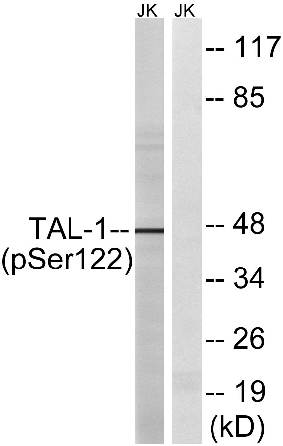
- Western blot analysis of lysates from Jurkat cells, treated with PMA 125ng/ml 30', using TAL-1 Antibody. The lane on the right is blocked with the synthesized peptide.
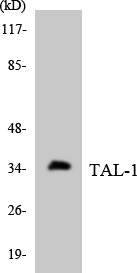
- Western blot analysis of the lysates from HT-29 cells using TAL-1 antibody.
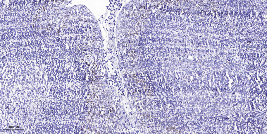
- Immunohistochemical analysis of paraffin-embedded human tonsil. 1, Antibody was diluted at 1:200(4° overnight). 2, Tris-EDTA,pH9.0 was used for antigen retrieval. 3,Secondary antibody was diluted at 1:200(room temperature, 45min).