- 首页
- 公司介绍
- 热门促销
-
全部产品
-
试剂盒
- |
-
一抗
- |
-
二抗
- |
-
蛋白
- |
-
免疫组化试剂
- |
-
WB 试剂
- PonceauS Staining Solution
- PBST Washing Buffer, 10X
- 1.5M Tris-HCl Buffer, pH8.8
- 1M Tris-HCl Buffer, pH6.8
- 10% SDS Solution
- Prestained Protein Marker
- TBST Washing Buffer, 10X
- SDS PAGE Loading Buffer, 5X
- Stripping Buffered Solution
- Tris Buffer, pH7.4, 10X
- Total Protein Extraction Kit
- Running Buffer, 10X
- Transfer Buffer, 10X
- 30% Acr-Bis(29:1) Solution
- Tris电泳液速溶颗粒
- PBS(1X, premixed powder)
- TBS(1X, premixed powder)
- 快速封闭液
- 转膜液速溶颗粒
- Chemical reagents
- 公司新闻
- 营销网络
- 资源中心
- 联系我们
TTYH2 rabbit pAb
- 货号:YT7051
- 应用:WB
- 种属:Human;Mouse
- 免疫原:
- Synthesized peptide derived from human TTYH2 AA range: 319-369
- 特异性:
- This antibody detects endogenous levels of TTYH2 at Human/Mouse
- 组成:
- Liquid in PBS containing 50% glycerol, 0.5% BSA and 0.02% sodium azide.
- 来源:
- Polyclonal, Rabbit,IgG
- 纯化工艺:
- The antibody was affinity-purified from rabbit antiserum by affinity-chromatography using epitope-specific immunogen.
- 储存:
- -15°C to -25°C/1 year(Do not lower than -25°C)
- 背景:
- This gene encodes a member of the tweety family of proteins. Members of this family function as chloride anion channels. The encoded protein functions as a calcium(2+)-activated large conductance chloride(-) channel, and may play a role in kidney tumorigenesis. Two transcript variants encoding distinct isoforms have been identified for this gene. [provided by RefSeq, Jul 2008],
- 功能:
- function:Probable large-conductance Ca(2+)-activated chloride channel. May play a role in Ca(2+) signal transduction. May be involved in cell proliferation and cell aggregation.,similarity:Belongs to the tweety family.,tissue specificity:Expressed at higher level in brain and testis and at lower levels in heart, ovary, spleen and peripheral blood leukocytes. Up-regulated in 13 of 16 renal cell carcinoma samples examined. Up-regulated in colon carcinoma.,
- 细胞定位:
- Cell membrane ; Multi-pass membrane protein .
- 组织表达:
- Expressed at higher level in brain and testis and at lower levels in heart, ovary, spleen and peripheral blood leukocytes. Up-regulated in 13 of 16 renal cell carcinoma samples examined. Up-regulated in colon carcinoma.
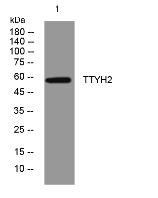
- Western blot analysis of lysates from 293T cells, primary antibody was diluted at 1:1000, 4°over night


