- 首页
- 公司介绍
- 热门促销
-
全部产品
-
试剂盒
- |
-
一抗
- |
-
二抗
- |
-
蛋白
- |
-
免疫组化试剂
- |
-
WB 试剂
- PonceauS Staining Solution
- PBST Washing Buffer, 10X
- 1.5M Tris-HCl Buffer, pH8.8
- 1M Tris-HCl Buffer, pH6.8
- 10% SDS Solution
- Prestained Protein Marker
- TBST Washing Buffer, 10X
- SDS PAGE Loading Buffer, 5X
- Stripping Buffered Solution
- Tris Buffer, pH7.4, 10X
- Total Protein Extraction Kit
- Running Buffer, 10X
- Transfer Buffer, 10X
- 30% Acr-Bis(29:1) Solution
- Tris电泳液速溶颗粒
- PBS(1X, premixed powder)
- TBS(1X, premixed powder)
- 快速封闭液
- 转膜液速溶颗粒
- Chemical reagents
- 公司新闻
- 营销网络
- 资源中心
- 联系我们
ASTL rabbit pAb
- 货号:YT6403
- 应用:WB
- 种属:Human;Mouse
- 免疫原:
- Synthesized peptide derived from human ASTL AA range: 115-165
- 特异性:
- This antibody detects endogenous levels of ASTL at Human/Mouse
- 组成:
- Liquid in PBS containing 50% glycerol, 0.5% BSA and 0.02% sodium azide.
- 来源:
- Polyclonal, Rabbit,IgG
- 纯化工艺:
- The antibody was affinity-purified from rabbit antiserum by affinity-chromatography using epitope-specific immunogen.
- 储存:
- -15°C to -25°C/1 year(Do not lower than -25°C)
- 功能:
- enzyme regulation:Inhibited by wide spectrum metalloproteinase inhibitor batimastat (BB-94). Also inhibited by EDTA.,similarity:Belongs to the peptidase M12A family.,tissue specificity:Expressed in promyelocytic leukemia HL-60 cells, Burkitt's lymphoma Raji cells, and also in some ovarian carcinomas. Not detected in normal tissues.,
- 细胞定位:
- Cytoplasm . Cell membrane . Cytoplasmic vesicle, secretory vesicle, Cortical granule . Probably exocytosed from cortical granules during post-fertilization. Detected throughout the ooplasm of germinal vesicle stage oocytes in early bilaminar secondary follicles at postnatal (PN) day 3. Detected in the microvillar domain of the oolemma in arrested ovulated secondary oocytes and in the first polar body prior to fertilization. Upon fertilization, detected in the perivitelline space (PVS) and occasionally on the oolemma in 2-cell through morulae stages. Colocalizes with SPACA3 at the microvillar domain of the oolemma and in the perivitelline space (PVS). .
- 组织表达:
- Expressed in promyelocytic leukemia HL-60 cells, Burkitt's lymphoma Raji cells, and also in some ovarian carcinomas. Not detected in normal tissues.
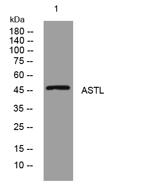
- Western blot analysis of lysates from HEK293 cells, primary antibody was diluted at 1:1000, 4°over night


