Cystatin A Polyclonal Antibody
- 货号:YT5876
- 应用:WB;IHC;IF;ELISA
- 种属:Human;Rat;Mouse;
- 蛋白名称:
- Cystatin-A (Cystatin-AS) (Stefin-A)
- 免疫原:
- Synthetic peptide from human protein at AA range: 60-98
- 特异性:
- The antibody detects endogenous Cystatin A
- 组成:
- Liquid in PBS containing 50% glycerol, 0.5% BSA and 0.02% sodium azide.
- 来源:
- Polyclonal, Rabbit,IgG
- 稀释:
- WB 1:500-2000,IHC 1:500-200, ELISA 1:10000-20000. IF 1:50-200
- 纯化工艺:
- The antibody was affinity-purified from rabbit antiserum by affinity-chromatography using epitope-specific immunogen.
- 储存:
- -15°C to -25°C/1 year(Do not lower than -25°C)
- 其他名称:
- Cystatin-A (Cystatin-AS;Stefin-A)
- 背景:
- The cystatin superfamily encompasses proteins that contain multiple cystatin-like sequences. Some of the members are active cysteine protease inhibitors, while others have lost or perhaps never acquired this inhibitory activity. There are three inhibitory families in the superfamily, including the type 1 cystatins (stefins), type 2 cystatins, and kininogens. This gene encodes a stefin that functions as a cysteine protease inhibitor, forming tight complexes with papain and the cathepsins B, H, and L. The protein is one of the precursor proteins of cornified cell envelope in keratinocytes and plays a role in epidermal development and maintenance. Stefins have been proposed as prognostic and diagnostic tools for cancer. [provided by RefSeq, Jul 2008],
- 功能:
- function:This is an intracellular thiol proteinase inhibitor.,similarity:Belongs to the cystatin family.,
- 组织表达:
- Expressed in the skin throughout the epidermis.

- Western blot analysis of 293T mouse-kidney lysate, antibody was diluted at 1000. Secondary antibody(catalog#:RS0002) was diluted at 1:20000
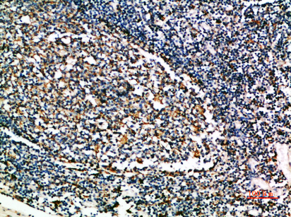
- Immunohistochemical analysis of paraffin-embedded human-tonsil, antibody was diluted at 1:200


