Annexin I Polyclonal Antibody
- 货号:YT5872
- 应用:IF;WB;IHC;ELISA
- 种属:Human;Rat;Mouse;
- 蛋白名称:
- Annexin A1 (Annexin I) (Annexin-1) (Calpactin II) (Calpactin-2) (Chromobindin-9) (Lipocortin I) (Phospholipase A2 inhibitory protein) (p35)
- 免疫原:
- Synthetic peptide from human protein at AA range: 130-180
- 特异性:
- The antibody detects endogenous Annexin I
- 组成:
- Liquid in PBS containing 50% glycerol, 0.5% BSA and 0.02% sodium azide.
- 来源:
- Polyclonal, Rabbit,IgG
- 稀释:
- IF 1:50-200 WB 1:500-2000,IHC 1:500-200, ELISA 1:10000-20000
- 纯化工艺:
- The antibody was affinity-purified from rabbit antiserum by affinity-chromatography using epitope-specific immunogen.
- 储存:
- -15°C to -25°C/1 year(Do not lower than -25°C)
- 其他名称:
- Annexin A1 (Annexin I) (Annexin-1) (Calpactin II) (Calpactin-2) (Chromobindin-9) (Lipocortin I) (Phospholipase A2 inhibitory protein) (p35)
- 背景:
- This gene encodes a membrane-localized protein that binds phospholipids. This protein inhibits phospholipase A2 and has anti-inflammatory activity. Loss of function or expression of this gene has been detected in multiple tumors. [provided by RefSeq, Dec 2014],
- 功能:
- domain:A pair of annexin repeats may form one binding site for calcium and phospholipid.,function:Calcium/phospholipid-binding protein which promotes membrane fusion and is involved in exocytosis. This protein regulates phospholipase A2 activity. It seems to bind from two to four calcium ions with high affinity.,PTM:Phosphorylated by protein kinase C, epidermal growth factor receptor/kinase and TRPM7. Phosphorylation results in loss of the inhibitory activity.,similarity:Belongs to the annexin family.,similarity:Contains 1 annexin repeat.,similarity:Contains 2 annexin repeats.,similarity:Contains 4 annexin repeats.,subcellular location:Found in the cilium, nucleus and basolateral cell membrane of ciliated cells in the tracheal endothelium (By similarity). Found in the cytoplasm of type II pneumocytes and alveolar macrophages.,subunit:Homodimer in placenta (20%); linked by transglutamylat
- 细胞定位:
- Nucleus . Cytoplasm . Cell projection, cilium . Cell membrane . Membrane ; Peripheral membrane protein . Endosome membrane ; Peripheral membrane protein . Basolateral cell membrane . Apical cell membrane . Lateral cell membrane . Secreted . Secreted, extracellular space . Cell membrane ; Peripheral membrane protein ; Extracellular side . Secreted, extracellular exosome . Cytoplasmic vesicle, secretory vesicle lumen . Cell projection, phagocytic cup . Early endosome . Cytoplasmic vesicle membrane ; Peripheral membrane protein . Secreted, at least in part via exosomes and other secretory vesicles. Detected in exosomes and other extracellular vesicles (PubMed:25664854). Alternatively, the secretion is dependent on protein unfolding and facilitated by the cargo receptor TMED10; it results in t
- 组织表达:
- Detected in resting neutrophils (PubMed:10772777). Detected in peripheral blood T-cells (PubMed:17008549). Detected in extracellular vesicles in blood serum from patients with inflammatory bowel disease, but not in serum from healthy donors (PubMed:25664854). Detected in placenta (at protein level) (PubMed:2532504). Detected in liver.
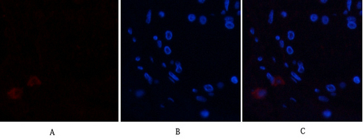
- Immunofluorescence analysis of human-breast tissue. 1,Annexin I Polyclonal Antibody(red) was diluted at 1:200(4°C,overnight). 2, Cy3 labled Secondary antibody was diluted at 1:300(room temperature, 50min).3, Picture B: DAPI(blue) 10min. Picture A:Target. Picture B: DAPI. Picture C: merge of A+B
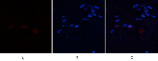
- Immunofluorescence analysis of human-breast tissue. 1,Annexin I Polyclonal Antibody(red) was diluted at 1:200(4°C,overnight). 2, Cy3 labled Secondary antibody was diluted at 1:300(room temperature, 50min).3, Picture B: DAPI(blue) 10min. Picture A:Target. Picture B: DAPI. Picture C: merge of A+B
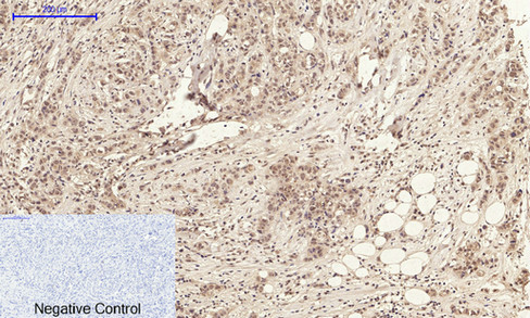
- Immunohistochemical analysis of paraffin-embedded Human-breast-cancer tissue. 1,Annexin I Polyclonal Antibody was diluted at 1:200(4°C,overnight). 2, Sodium citrate pH 6.0 was used for antibody retrieval(>98°C,20min). 3,Secondary antibody was diluted at 1:200(room tempeRature, 30min). Negative control was used by secondary antibody only.
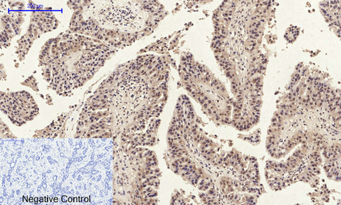
- Immunohistochemical analysis of paraffin-embedded Human-liver-cancer tissue. 1,Annexin I Polyclonal Antibody was diluted at 1:200(4°C,overnight). 2, Sodium citrate pH 6.0 was used for antibody retrieval(>98°C,20min). 3,Secondary antibody was diluted at 1:200(room tempeRature, 30min). Negative control was used by secondary antibody only.
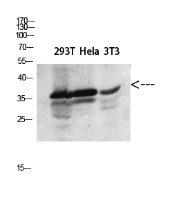
- Western blot analysis of 293T Hela lysate, antibody was diluted at 2000. Secondary antibody(catalog#:RS0002) was diluted at 1:20000
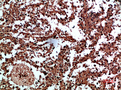
- Immunohistochemical analysis of paraffin-embedded human-spleen, antibody was diluted at 1:200


