VN2R1P Polyclonal Antibody
- 货号:YT4886
- 应用:WB;IF;ELISA
- 种属:Human;Rat;Mouse;
- 蛋白名称:
- Putative calcium-sensing receptor-like 1
- 免疫原:
- The antiserum was produced against synthesized peptide derived from human CASRL1. AA range:400-449
- 特异性:
- VN2R1P Polyclonal Antibody detects endogenous levels of VN2R1P protein.
- 组成:
- Liquid in PBS containing 50% glycerol, 0.5% BSA and 0.02% sodium azide.
- 来源:
- Polyclonal, Rabbit,IgG
- 稀释:
- WB 1:500 - 1:2000. IF 1:200 - 1:1000. ELISA: 1:10000. Not yet tested in other applications.
- 纯化工艺:
- The antibody was affinity-purified from rabbit antiserum by affinity-chromatography using epitope-specific immunogen.
- 储存:
- -15°C to -25°C/1 year(Do not lower than -25°C)
- 背景:
- The CARL-1 monoclonal antibody reacts with human TWEAK, a type II transmembrane TNF superfamily member with high identity to TNF in its extracellular portion. TWEAK transcript is expressed broadly in many adult and fetal tissues, however, the staining of human peripheral blood mononuclear cells with monoclonal antibodies shows a more restricted pattern. While freshly isolated PBMCs do not express detectable levels of TWEAK on their surface, IFN-gamma-stimulated blood monocytes rapidly upregulate TWEAK surface expression. TWEAK is expressed as membrane bound and secreted forms. Interaction of TWEAK with its counter-receptor promotes secretion of IL-8, activation of NF-kappaB, proliferation of endothelial cells, and apoptosis in a number of human cell lines. Initially, DR3 was thought to be a receptor for TWEAK, but further studies have shown that TWEAK could induce apoptosis via receptors distinct from DR3. While TWEAK exhibits overlapping signaling functions to TNF, it is generally less effective in inducing apoptosis, giving rise to its name, TNF-like weak inducer of apoptosis. For detection of human TWEAK by sandwich ELISA, a combination of purified CARL-2 for capture and biotinylated CARL-1 for detection is recommended.

- Immunofluorescence analysis of A549 cells, using CSRL1 Antibody. The picture on the right is blocked with the synthesized peptide.

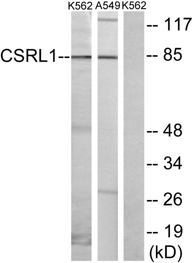
- Western blot analysis of lysates from K562 cells and A549 cells, using CSRL1 Antibody. The lane on the right is blocked with the synthesized peptide.


