TRH-R1 Polyclonal Antibody
- 货号:YT4734
- 应用:WB;IF;ELISA
- 种属:Human;Mouse;Rat;Monkey
- 简介:
- >>Calcium signaling pathway;>>Neuroactive ligand-receptor interaction
- 蛋白名称:
- Thyrotropin-releasing hormone receptor
- 免疫原:
- The antiserum was produced against synthesized peptide derived from human TRHR. AA range:195-244
- 特异性:
- TRH-R1 Polyclonal Antibody detects endogenous levels of TRH-R1 protein.
- 组成:
- Liquid in PBS containing 50% glycerol, 0.5% BSA and 0.02% sodium azide.
- 来源:
- Polyclonal, Rabbit,IgG
- 稀释:
- WB 1:500 - 1:2000. IF 1:200 - 1:1000. ELISA: 1:20000. Not yet tested in other applications.
- 纯化工艺:
- The antibody was affinity-purified from rabbit antiserum by affinity-chromatography using epitope-specific immunogen.
- 储存:
- -15°C to -25°C/1 year(Do not lower than -25°C)
- 其他名称:
- TRHR;Thyrotropin-releasing hormone receptor;TRH-R;Thyroliberin receptor
- 背景:
- This gene encodes a G protein-coupled receptor for thyrotropin-releasing hormone (TRH). Upon binding to TRH, this receptor activates the inositol phospholipid-calcium-protein kinase C transduction pathway. Mutations in this gene have been associated with generalized thyrotropin-releasing hormone resistance. [provided by RefSeq, Sep 2011],
- 功能:
- function:Receptor for thyrotropin-releasing hormone. This receptor is mediated by G proteins which activate a phosphatidylinositol-calcium second messenger system.,similarity:Belongs to the G-protein coupled receptor 1 family.,
- 细胞定位:
- Cell membrane ; Multi-pass membrane protein .
- 组织表达:
- Brain,Pituitary,Placenta,

- Immunofluorescence analysis of A549 cells, using TRHR Antibody. The picture on the right is blocked with the synthesized peptide.
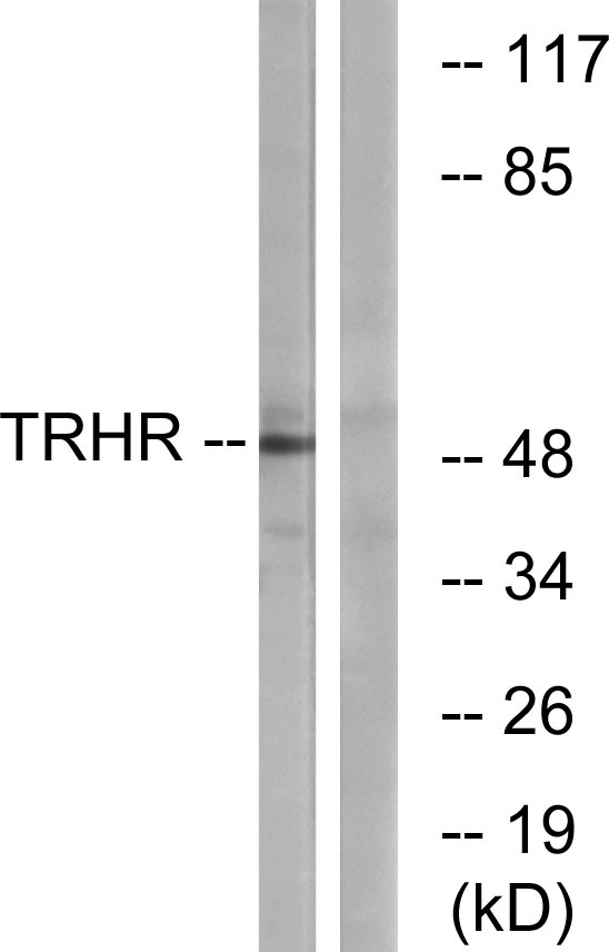
- Western blot analysis of lysates from COS7 cells, using TRHR Antibody. The lane on the right is blocked with the synthesized peptide.
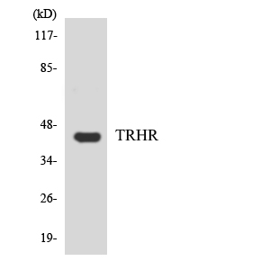
- Western blot analysis of the lysates from HepG2 cells using TRHR antibody.


