Rho H Polyclonal Antibody
- 货号:YT4083
- 应用:WB;IHC;IF;ELISA
- 种属:Human;Mouse;Rat
- 简介:
- >>Leukocyte transendothelial migration;>>Salmonella infection
- 蛋白名称:
- Rho-related GTP-binding protein RhoH
- 免疫原:
- The antiserum was produced against synthesized peptide derived from human RhoH. AA range:141-190
- 特异性:
- Rho H Polyclonal Antibody detects endogenous levels of Rho H protein.
- 组成:
- Liquid in PBS containing 50% glycerol, 0.5% BSA and 0.02% sodium azide.
- 来源:
- Polyclonal, Rabbit,IgG
- 稀释:
- WB 1:500 - 1:2000. IHC 1:100 - 1:300. IF 1:200 - 1:1000. ELISA: 1:20000. Not yet tested in other applications.
- 纯化工艺:
- The antibody was affinity-purified from rabbit antiserum by affinity-chromatography using epitope-specific immunogen.
- 储存:
- -15°C to -25°C/1 year(Do not lower than -25°C)
- 其他名称:
- RHOH;ARHH;TTF;Rho-related GTP-binding protein RhoH;GTP-binding protein TTF;Translocation three four protein
- 背景:
- The protein encoded by this gene is a member of the Ras superfamily of guanosine triphosphate (GTP)-metabolizing enzymes. The encoded protein is expressed in hematopoietic cells, where it functions as a negative regulator of cell growth and survival. This gene may be hypermutated or misexpressed in leukemias and lymphomas. Chromosomal translocations in non-Hodgkin's lymphoma occur between this locus and B-cell CLL/lymphoma 6 (BCL6) on chromosome 3, leading to the production of fusion transcripts. Alternative splicing in the 5' untranslated region results in multiple transcript variants that encode the same protein. [provided by RefSeq, May 2013],
- 功能:
- disease:A chromosomal aberration involving RHOH is found in a non-Hodgkin lymphoma cell line. Translocation t(3;4)(q27;p11) with BCL6.,similarity:Belongs to the small GTPase superfamily. Rho family.,tissue specificity:Transcribed only in hemopoietic cells.,
- 细胞定位:
- Cytoplasm . Cell membrane ; Lipid-anchor ; Cytoplasmic side . Colocalizes together with ZAP70 in the immunological synapse. .
- 组织表达:
- Expressed only in hematopoietic cells. Present at very high levels in the thymus, less abundant in the spleen, and least abundant in the bone marrow. Expressed at a higher level in the TH1 subtype of T-helper cells than in the TH2 subpopulation. Expressed in neutrophils under inflammatory conditions, such as cystic fibrosis, ulcerative colitis and appendicitis.

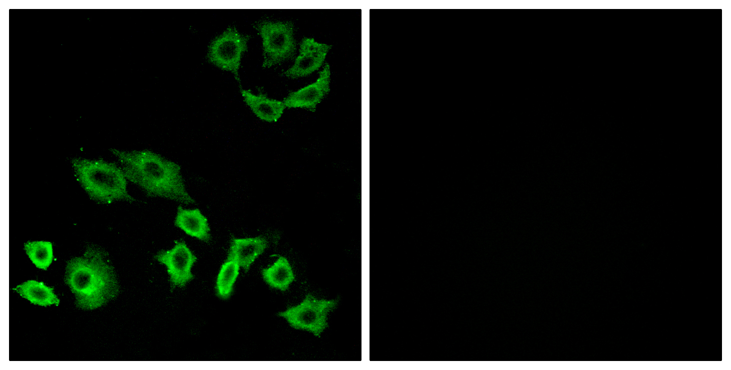
- Immunofluorescence analysis of A549 cells, using RhoH Antibody. The picture on the right is blocked with the synthesized peptide.

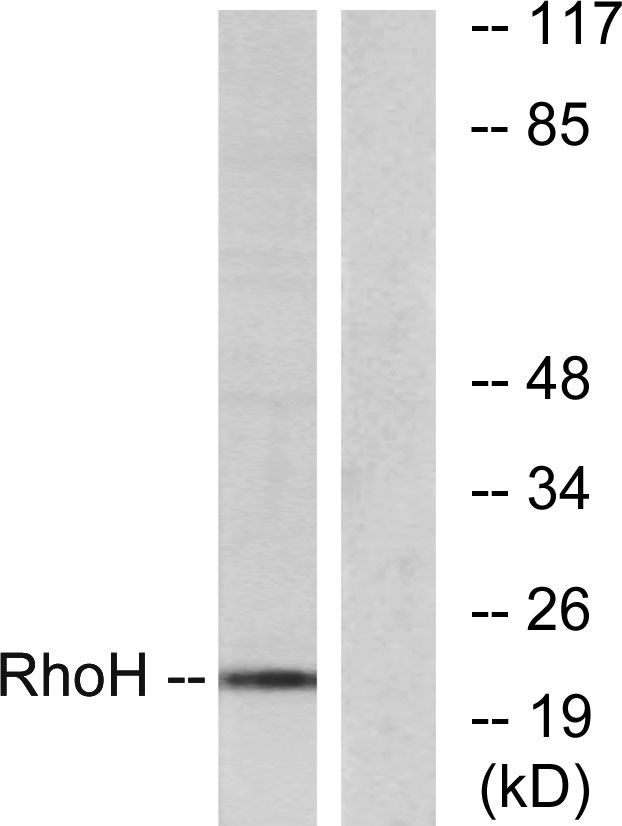
- Western blot analysis of lysates from HT-29 cells, using RhoH Antibody. The lane on the right is blocked with the synthesized peptide.

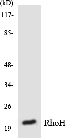
- Western blot analysis of the lysates from RAW264.7cells using RhoH antibody.

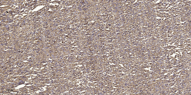
- Immunohistochemical analysis of paraffin-embedded human small intestinal carcinoma tissue. 1,primary Antibody was diluted at 1:200(4° overnight). 2, Sodium citrate pH 6.0 was used for antigen retrieval(>98°C,20min). 3,Secondary antibody was diluted at 1:200


