- 首页
- 公司介绍
- 热门促销
-
全部产品
-
试剂盒
- |
-
一抗
- |
-
二抗
- |
-
蛋白
- |
-
免疫组化试剂
- |
-
WB 试剂
- PonceauS Staining Solution
- PBST Washing Buffer, 10X
- 1.5M Tris-HCl Buffer, pH8.8
- 1M Tris-HCl Buffer, pH6.8
- 10% SDS Solution
- Prestained Protein Marker
- TBST Washing Buffer, 10X
- SDS PAGE Loading Buffer, 5X
- Stripping Buffered Solution
- Tris Buffer, pH7.4, 10X
- Total Protein Extraction Kit
- Running Buffer, 10X
- Transfer Buffer, 10X
- 30% Acr-Bis(29:1) Solution
- Tris电泳液速溶颗粒
- PBS(1X, premixed powder)
- TBS(1X, premixed powder)
- 快速封闭液
- 转膜液速溶颗粒
- Chemical reagents
- 公司新闻
- 营销网络
- 资源中心
- 联系我们
MDM1 Polyclonal Antibody
- 货号:YT2689
- 应用:WB;ELISA
- 种属:Human;Mouse;Rat
- 蛋白名称:
- Nuclear protein MDM1
- 免疫原:
- The antiserum was produced against synthesized peptide derived from human MDM1. AA range:665-714
- 特异性:
- MDM1 Polyclonal Antibody detects endogenous levels of MDM1 protein.
- 组成:
- Liquid in PBS containing 50% glycerol, 0.5% BSA and 0.02% sodium azide.
- 来源:
- Polyclonal, Rabbit,IgG
- 稀释:
- WB 1:500 - 1:2000. ELISA: 1:5000. Not yet tested in other applications.
- 纯化工艺:
- The antibody was affinity-purified from rabbit antiserum by affinity-chromatography using epitope-specific immunogen.
- 储存:
- -15°C to -25°C/1 year(Do not lower than -25°C)
- 其他名称:
- MDM1;Nuclear protein MDM1
- 背景:
- This gene encodes a nuclear protein similar to the mouse double minute 1 protein. The mouse gene is located in double minute (DM) chromatin particles, is amplified in the mouse transformed 3T3 cell line, and the encoded protein is able to bind to p53. Alternatively spliced transcript variants encoding different isoforms have been found for this gene. [provided by RefSeq, Mar 2011],
- 功能:
- similarity:Belongs to the MDM1 family.,
- 细胞定位:
- Nucleus . Cytoplasm, cytoskeleton, microtubule organizing center, centrosome . Cytoplasm, cytoskeleton, microtubule organizing center, centrosome, centriole . Localizes to the centriole lumen. .
- 组织表达:
- Brain,Epithelium,Prostate,Testis,Trachea,
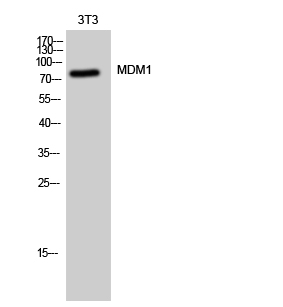
- Western Blot analysis of 3T3 cells using MDM1 Polyclonal Antibody cells nucleus extracted by Minute TM Cytoplasmic and Nuclear Fractionation kit (SC-003,Inventbiotech,MN,USA).

- Western blot analysis of MDM1 Antibody. The lane on the right is blocked with the MDM1 peptide.
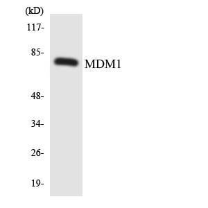
- Western blot analysis of the lysates from HepG2 cells using MDM1 antibody.


