- 首页
- 公司介绍
- 热门促销
-
全部产品
-
试剂盒
- |
-
一抗
- |
-
二抗
- |
-
蛋白
- |
-
免疫组化试剂
- |
-
WB 试剂
- PonceauS Staining Solution
- PBST Washing Buffer, 10X
- 1.5M Tris-HCl Buffer, pH8.8
- 1M Tris-HCl Buffer, pH6.8
- 10% SDS Solution
- Prestained Protein Marker
- TBST Washing Buffer, 10X
- SDS PAGE Loading Buffer, 5X
- Stripping Buffered Solution
- Tris Buffer, pH7.4, 10X
- Total Protein Extraction Kit
- Running Buffer, 10X
- Transfer Buffer, 10X
- 30% Acr-Bis(29:1) Solution
- Tris电泳液速溶颗粒
- PBS(1X, premixed powder)
- TBS(1X, premixed powder)
- 快速封闭液
- 转膜液速溶颗粒
- Chemical reagents
- 公司新闻
- 营销网络
- 资源中心
- 联系我们
ILK Polyclonal Antibody
- 货号:YT2346
- 应用:WB;IHC;IF;ELISA
- 种属:Human;Mouse;Rat
- 简介:
- >>PPAR signaling pathway;>>Axon guidance;>>Focal adhesion;>>Bacterial invasion of epithelial cells;>>Shigellosis;>>Endometrial cancer
- 蛋白名称:
- Integrin-linked protein kinase
- 免疫原:
- The antiserum was produced against synthesized peptide derived from human ILK. AA range:212-261
- 特异性:
- ILK Polyclonal Antibody detects endogenous levels of ILK protein.
- 组成:
- Liquid in PBS containing 50% glycerol, 0.5% BSA and 0.02% sodium azide.
- 来源:
- Polyclonal, Rabbit,IgG
- 稀释:
- WB 1:500 - 1:2000. IHC 1:100 - 1:300. ELISA: 1:5000.. IF 1:50-200
- 纯化工艺:
- The antibody was affinity-purified from rabbit antiserum by affinity-chromatography using epitope-specific immunogen.
- 储存:
- -15°C to -25°C/1 year(Do not lower than -25°C)
- 其他名称:
- ILK;ILK1;ILK2;Integrin-linked protein kinase;59 kDa serine/threonine-protein kinase;ILK-1;ILK-2;p59ILK
- 背景:
- This gene encodes a protein with a kinase-like domain and four ankyrin-like repeats. The encoded protein associates at the cell membrane with the cytoplasmic domain of beta integrins, where it regulates integrin-mediated signal transduction. Activity of this protein is important in the epithelial to mesenchymal transition, and over-expression of this gene is implicated in tumor growth and metastasis. Alternative splicing results in multiple transcript variants. [provided by RefSeq, Jun 2013],
- 功能:
- catalytic activity:ATP + a protein = ADP + a phosphoprotein.,domain:A PH-like domain is involved in phosphatidylinositol phosphate binding.,enzyme regulation:Stimulated rapidly but transiently by both cell fibronectin interactions, as well as by insulin, in a PI3-K-dependent manner, likely via the binding of PtdIns(3,4,5)P3 with a PH-like domain of ILK.,function:Receptor-proximal protein kinase regulating integrin-mediated signal transduction. May act as a mediator of inside-out integrin signaling. Focal adhesion protein part of the complex ILK-PINCH. This complex is considered to be one of the convergence points of integrin- and growth factor-signaling pathway. Could be implicated in mediating cell architecture, adhesion to integrin substrates and anchorage-dependent growth in epithelial cells. Phosphorylates beta-1 and beta-3 integrin subunit on serine and threonine residues, but also
- 细胞定位:
- Cell junction, focal adhesion . Cell membrane; Peripheral membrane protein; Cytoplasmic side . Cell projection, lamellipodium . Cytoplasm, myofibril, sarcomere .
- 组织表达:
- Highly expressed in heart followed by skeletal muscle, pancreas and kidney. Weakly expressed in placenta, lung and liver.
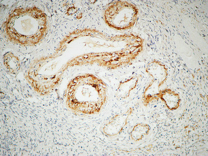
- Immunohistochemical analysis of paraffin-embedded Human ovary. 1, Antibody was diluted at 1:200(4° overnight). 2, High-pressure and temperature EDTA, pH8.0 was used for antigen retrieval. 3,Secondary antibody was diluted at 1:200(room temperature, 30min).
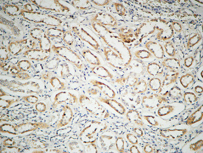
- Immunohistochemical analysis of paraffin-embedded Human kidney. 1, Antibody was diluted at 1:100(4° overnight). 2, High-pressure and temperature EDTA, pH8.0 was used for antigen retrieval. 3,Secondary antibody was diluted at 1:200(room temperature, 30min).
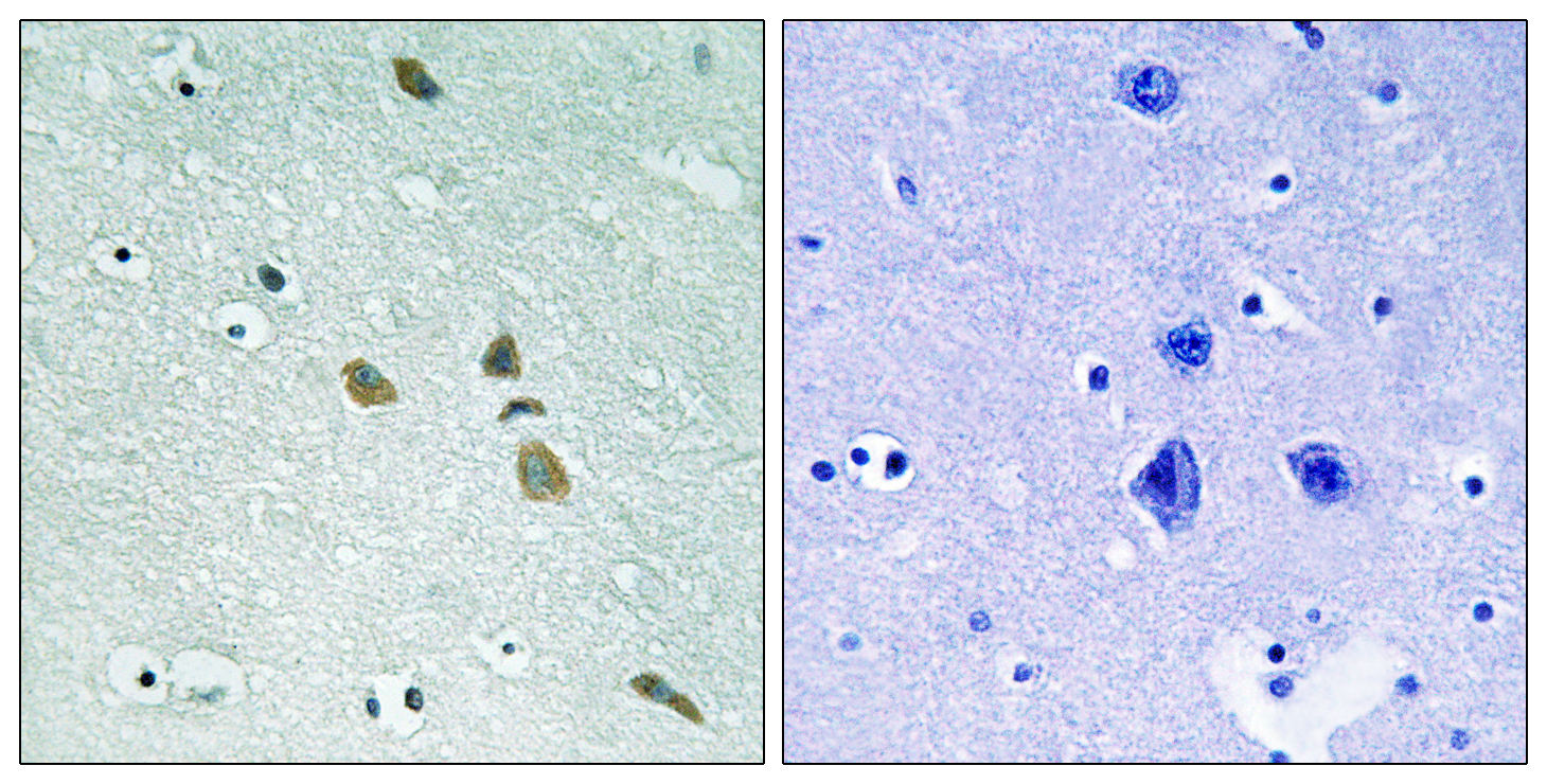
- Immunohistochemistry analysis of paraffin-embedded human brain tissue, using ILK Antibody. The picture on the right is blocked with the synthesized peptide.
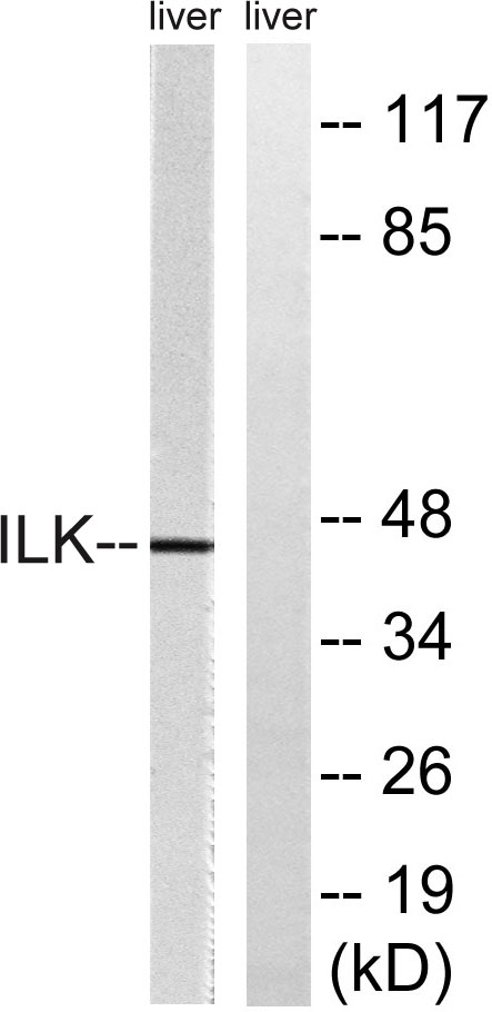
- Western blot analysis of lysates from rat liver cells, using ILK Antibody. The lane on the right is blocked with the synthesized peptide.
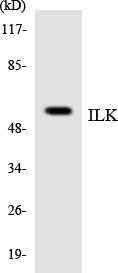
- Western blot analysis of the lysates from HUVECcells using ILK antibody.


