C-TAK1 Polyclonal Antibody
- 货号:YT1147
- 应用:WB;IHC;IF;ELISA
- 种属:Human;Mouse;Rat
- 蛋白名称:
- MAP/microtubule affinity-regulating kinase 3
- 免疫原:
- The antiserum was produced against synthesized peptide derived from human MARK3. AA range:1-50
- 特异性:
- C-TAK1 Polyclonal Antibody detects endogenous levels of C-TAK1 protein.
- 组成:
- Liquid in PBS containing 50% glycerol, 0.5% BSA and 0.02% sodium azide.
- 来源:
- Polyclonal, Rabbit,IgG
- 稀释:
- WB 1:500 - 1:2000. IHC 1:100 - 1:300. IF 1:200 - 1:1000. ELISA: 1:40000. Not yet tested in other applications.
- 纯化工艺:
- The antibody was affinity-purified from rabbit antiserum by affinity-chromatography using epitope-specific immunogen.
- 储存:
- -15°C to -25°C/1 year(Do not lower than -25°C)
- 其他名称:
- MARK3;CTAK1;EMK2;MAP/microtubule affinity-regulating kinase 3;C-TAK1;cTAK1;Cdc25C-associated protein kinase 1;ELKL motif kinase 2;EMK-2;Protein kinase STK10;Ser/Thr protein kinase PAR-1;Par-1a;Serine/threonine-protein kinase p78
- 背景:
- microtubule affinity regulating kinase 3(MARK3) Homo sapiens The protein encoded by this gene is activated by phosphorylation and in turn is involved in the phosphorylation of tau proteins MAP2 and MAP4. Several transcript variants encoding different isoforms have been found for this gene. [provided by RefSeq, Oct 2011],
- 功能:
- catalytic activity:ATP + a protein = ADP + a phosphoprotein.,function:Involved in the specific phosphorylation of microtubule-associated proteins for tau, MAP2 and MAP4. Phosphorylates CDC25C on 'Ser-216'.,similarity:Belongs to the protein kinase superfamily. CAMK Ser/Thr protein kinase family. MARK subfamily.,similarity:Contains 1 KA1 (kinase-associated) domain.,similarity:Contains 1 protein kinase domain.,similarity:Contains 1 UBA domain.,tissue specificity:Ubiquitous.,
- 细胞定位:
- Cell membrane ; Peripheral membrane protein . Cell projection, dendrite . Cytoplasm .

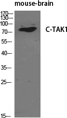
- Western Blot analysis of various cells using C-TAK1 Polyclonal Antibody diluted at 1:500
.jpg)
- Western Blot analysis of 293 cells using C-TAK1 Polyclonal Antibody diluted at 1:500

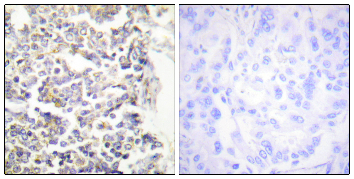
- Immunohistochemistry analysis of paraffin-embedded human lung carcinoma tissue, using MARK3 Antibody. The picture on the right is blocked with the synthesized peptide.

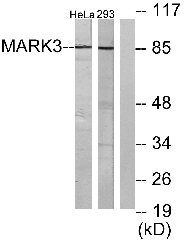
- Western blot analysis of lysates from HeLa and 293 cells, using MARK3 Antibody. The lane on the right is blocked with the synthesized peptide.

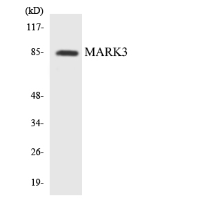
- Western blot analysis of the lysates from K562 cells using MARK3 antibody.

.jpg)


