Calmodulin Polyclonal Antibody
- 货号:YT0612
- 应用:WB;IHC;IF;ELISA
- 种属:Human;Mouse;Rat
- 免疫原:
- The antiserum was produced against synthesized peptide derived from human Calmodulin. AA range:46-95
- 特异性:
- Calmodulin Polyclonal Antibody detects endogenous levels of Calmodulin protein.
- 组成:
- Liquid in PBS containing 50% glycerol, 0.5% BSA and 0.02% sodium azide.
- 来源:
- Polyclonal, Rabbit,IgG
- 稀释:
- WB 1:500 - 1:2000. IHC 1:100 - 1:300. IF 1:200 - 1:1000. ELISA: 1:10000. Not yet tested in other applications.
- 纯化工艺:
- The antibody was affinity-purified from rabbit antiserum by affinity-chromatography using epitope-specific immunogen.
- 储存:
- -15°C to -25°C/1 year(Do not lower than -25°C)
- 其他名称:
- CALM1;CALM;CAM;CAM1;CALM2;CAM2;CAMB;CALM3;CALML2;CAM3;CAMC;CAMIII;Calmodulin;CaM
- 背景:
- This gene encodes a member of the EF-hand calcium-binding protein family. It is one of three genes which encode an identical calcium binding protein which is one of the four subunits of phosphorylase kinase. Two pseudogenes have been identified on chromosome 7 and X. Multiple transcript variants encoding different isoforms have been found for this gene.[provided by RefSeq, Oct 2009],
- 功能:
- function:Calmodulin mediates the control of a large number of enzymes and other proteins by Ca(2+). Among the enzymes to be stimulated by the calmodulin-Ca(2+) complex are a number of protein kinases and phosphatases. Together with CEP110 and centrin, is involved in a genetic pathway that regulates the centrosome cycle and progression through cytokinesis.,miscellaneous:This protein has four functional calcium-binding sites.,PTM:Phosphorylation results in a decreased activity.,PTM:Ubiquitination results in a strongly decreased activity.,similarity:Belongs to the calmodulin family.,similarity:Contains 4 EF-hand domains.,subcellular location:Distributed throughout the cell during interphase, but during mitosis becomes dramatically localized to the spindle poles and the spindle microtubules.,subunit:Interacts with MYO1C (By similarity). Interacts with CEP97, CEP110, TTN/titin and SRY.,
- 细胞定位:
- spindle pole,extracellular region,nucleus,nucleoplasm,cytoplasm,centrosome,cytosol,spindle microtubule,plasma membrane,voltage-gated potassium channel complex,sarcomere,growth cone,vesicle,calcium channel complex,G
- 组织表达:
- Blood,Brain,Cajal-Retzius cell,Fetal brain cortex,Lung,Lymph,Lymphoma,Muscle,Osteosarcoma,P
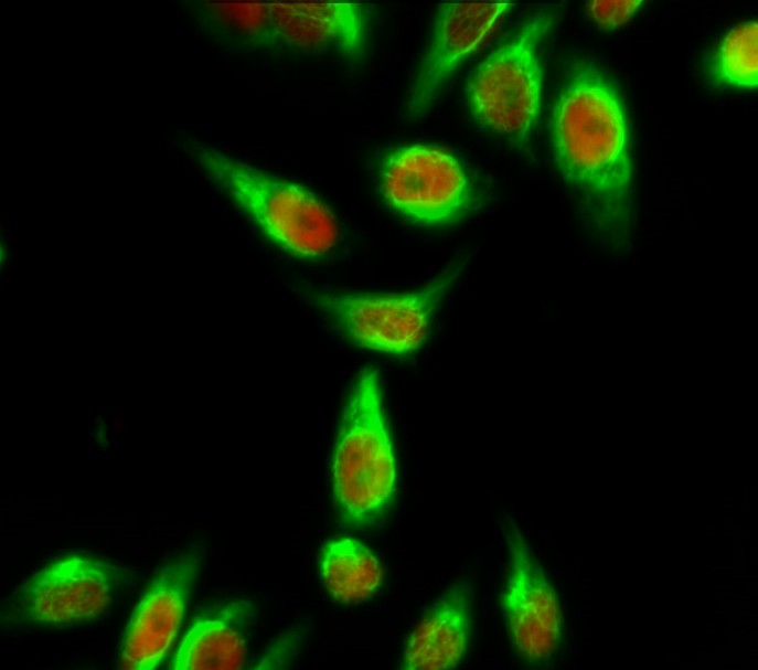
- Immunofluorescence analysis of Hela cell. 1,Calmodulin Polyclonal Antibody(green) was diluted at 1:200(4° overnight). (red) was diluted at 1:200(4° overnight). 2, Goat Anti Rabbit Alexa Fluor 488 Catalog:RS3211 was diluted at 1:1000(room temperature, 50min). Goat Anti Mouse Alexa Fluor 594 Catalog:RS3608 was diluted at 1:1000(room temperature, 50min).
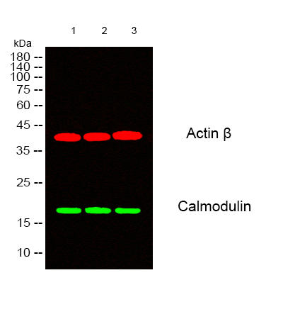
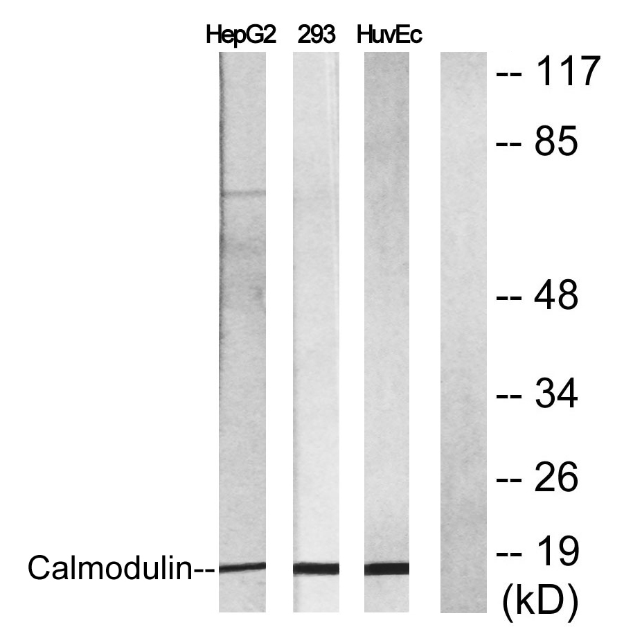
- Western blot analysis of lysates from 1) HuvEc, 2) HepG2, 3) 293 cells, (Green) primary antibody was diluted at 1:1000, 4°over night, secondary antibody(cat:RS23920)was diluted at 1:10000, 37° 1hour. (Red) Actin β Monoclonal Antibody(5B7) (cat:YM3028) antibody was diluted at 1:5000 as loading control, 4° over night,secondary antibody(cat:RS23710)was diluted at 1:10000, 37° 1hour.
- Western Blot analysis of various cells using Calmodulin Polyclonal Antibody diluted at 1:2000
.jpg)
- Western Blot analysis of HuvEc cells using Calmodulin Polyclonal Antibody diluted at 1:2000
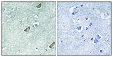
- Immunohistochemical analysis of paraffin-embedded Human brain. Antibody was diluted at 1:100(4° overnight). High-pressure and temperature Tris-EDTA,pH8.0 was used for antigen retrieval. Negetive contrl (right) obtaned from antibody was pre-absorbed by immunogen peptide.
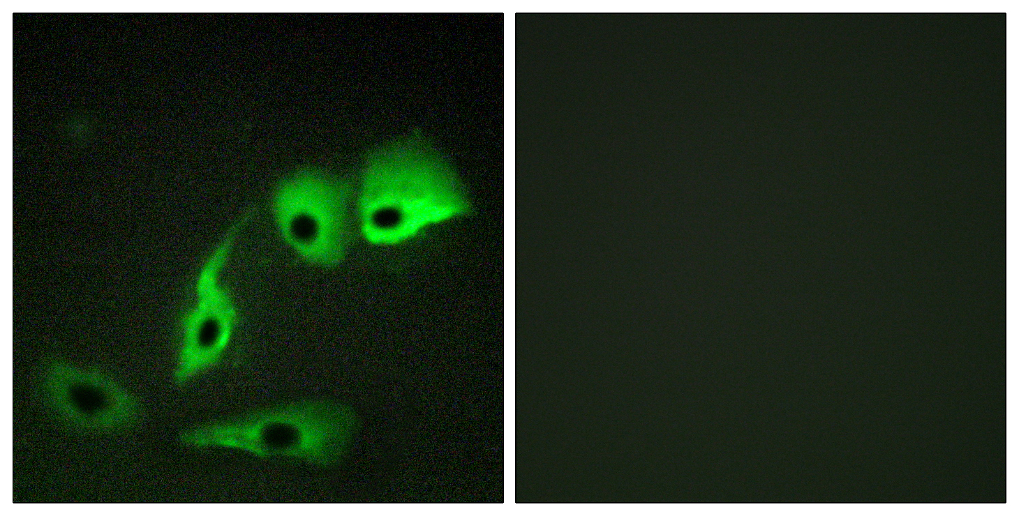
- Immunofluorescence analysis of HepG2 cells, using Calmodulin Antibody. The picture on the right is blocked with the synthesized peptide.
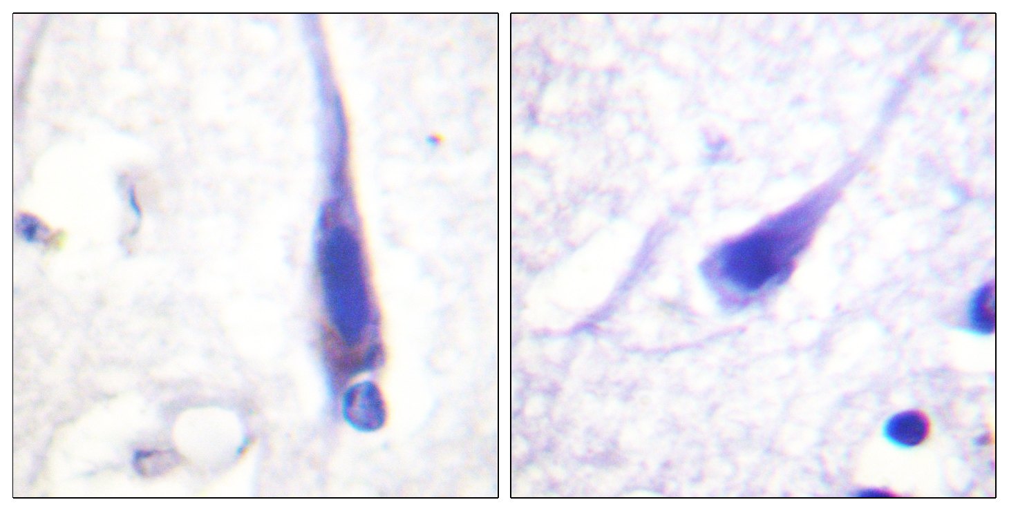
- Immunohistochemistry analysis of paraffin-embedded human brain tissue, using Calmodulin Antibody. The picture on the right is blocked with the synthesized peptide.
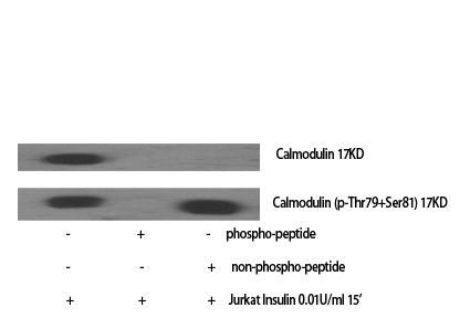
- Western blot analysis of lysates from NIH/3T3 cells, using Calmodulin Antibody. The lane on the right is blocked with the synthesized peptide.


