Cadherin-8 Polyclonal Antibody
- 货号:YT0603
- 应用:WB;IHC;IF;ELISA
- 种属:Human;Mouse;Rat
- 免疫原:
- The antiserum was produced against synthesized peptide derived from human CDH8. AA range:491-540
- 特异性:
- Cadherin-8 Polyclonal Antibody detects endogenous levels of Cadherin-8 protein.
- 组成:
- Liquid in PBS containing 50% glycerol, 0.5% BSA and 0.02% sodium azide.
- 来源:
- Polyclonal, Rabbit,IgG
- 稀释:
- WB 1:500 - 1:2000. IHC 1:100 - 1:300. IF 1:200 - 1:1000. ELISA: 1:20000. Not yet tested in other applications.
- 纯化工艺:
- The antibody was affinity-purified from rabbit antiserum by affinity-chromatography using epitope-specific immunogen.
- 储存:
- -15°C to -25°C/1 year(Do not lower than -25°C)
- 背景:
- This gene encodes a type II classical cadherin from the cadherin superfamily, integral membrane proteins that mediate calcium-dependent cell-cell adhesion. Mature cadherin proteins are composed of a large N-terminal extracellular domain, a single membrane-spanning domain, and a small, highly conserved C-terminal cytoplasmic domain. The extracellular domain consists of 5 subdomains, each containing a cadherin motif, and appears to determine the specificity of the protein's homophilic cell adhesion activity. Type II (atypical) cadherins are defined based on their lack of a HAV cell adhesion recognition sequence specific to type I cadherins. This particular cadherin is expressed in brain and is putatively involved in synaptic adhesion, axon outgrowth and guidance. [provided by RefSeq, Jul 2008],
- 功能:
- function:Cadherins are calcium dependent cell adhesion proteins. They preferentially interact with themselves in a homophilic manner in connecting cells; cadherins may thus contribute to the sorting of heterogeneous cell types.,similarity:Contains 5 cadherin domains.,tissue specificity:Mainly expressed in brain. Found in certain nerve cell lines, such as retinoblasts, glioma cells and neuroblasts.,
- 细胞定位:
- Cell membrane; Single-pass type I membrane protein.
- 组织表达:
- Mainly expressed in brain. Found in certain nerve cell lines, such as retinoblasts, glioma cells and neuroblasts.
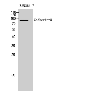
- Western Blot analysis of RAW264.7 cells using Cadherin-8 Polyclonal Antibody
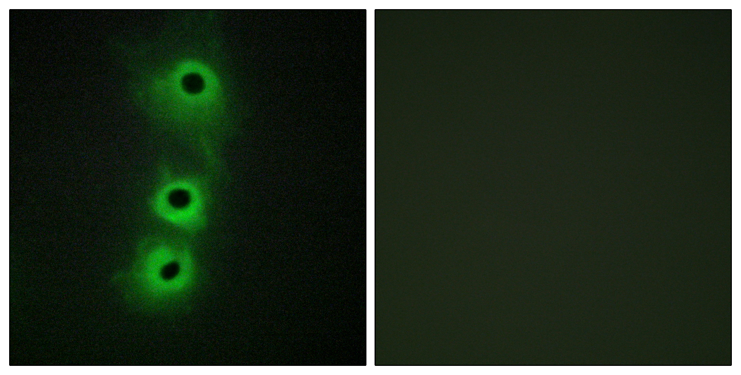
- Immunofluorescence analysis of COS7 cells, using CDH8 Antibody. The picture on the right is blocked with the synthesized peptide.
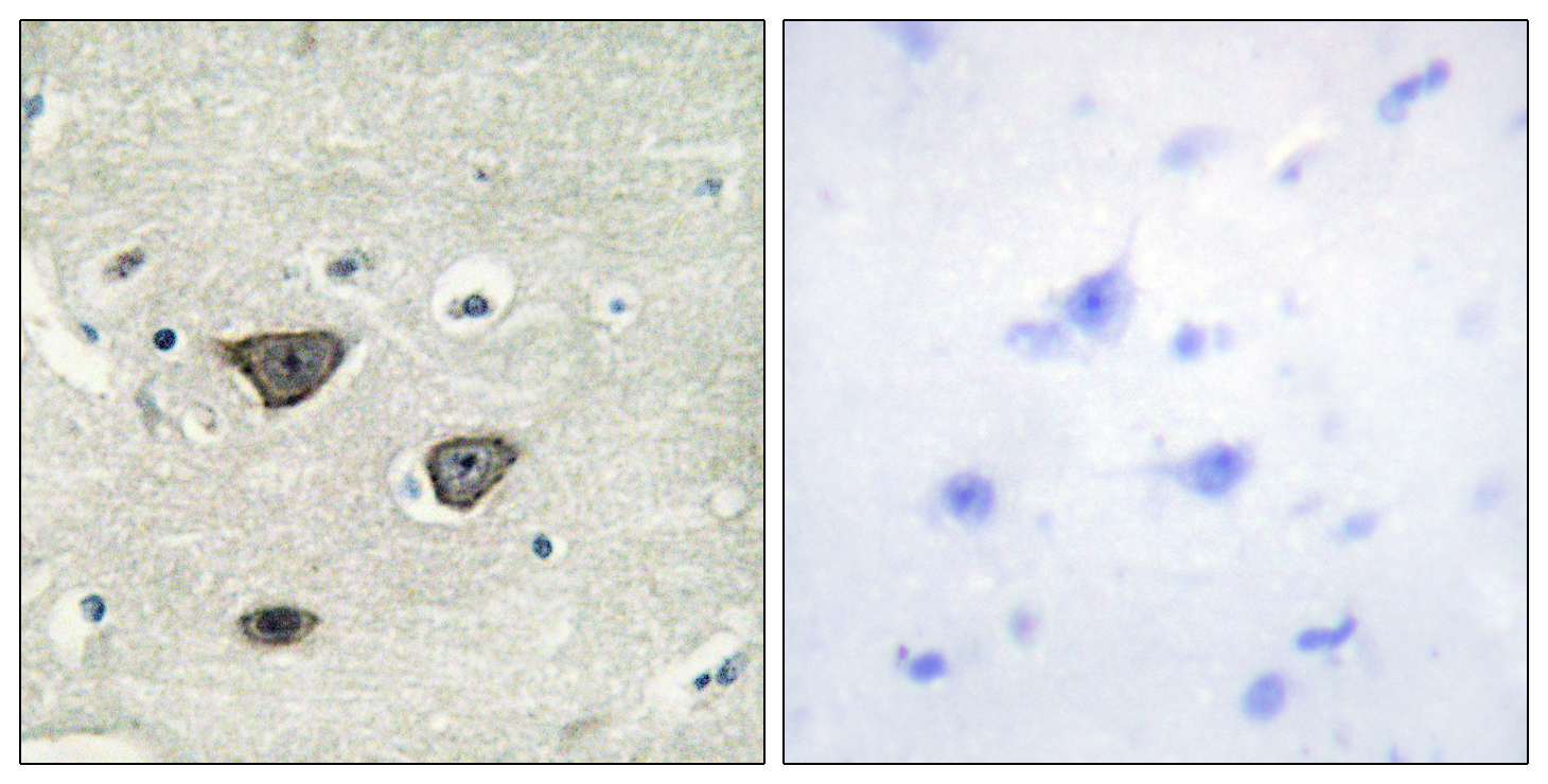
- Immunohistochemistry analysis of paraffin-embedded human brain tissue, using CDH8 Antibody. The picture on the right is blocked with the synthesized peptide.
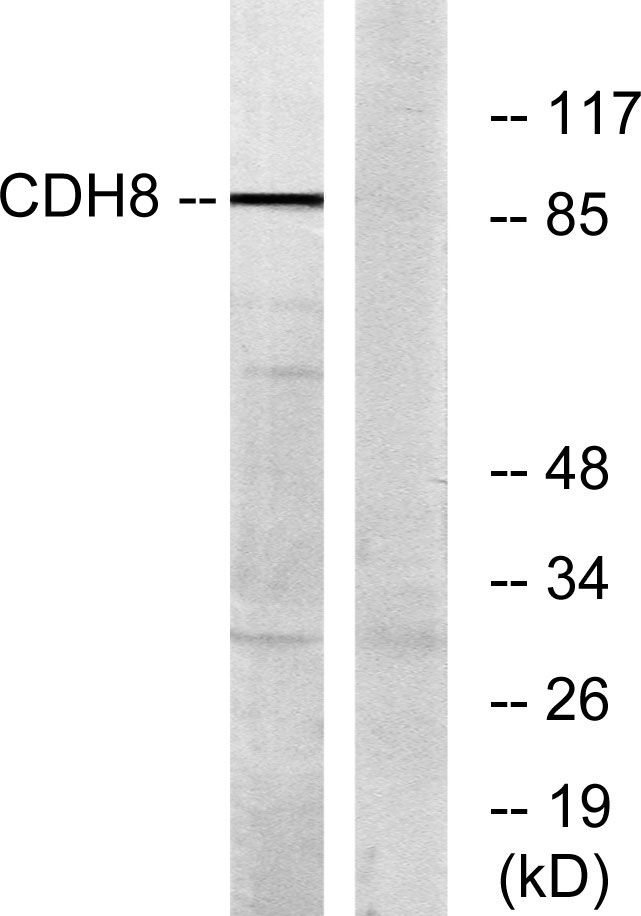
- Western blot analysis of lysates from RAW264.7 cells, using CDH8 Antibody. The lane on the right is blocked with the synthesized peptide.


