Cadherin-19 Polyclonal Antibody
- 货号:YT0596
- 应用:WB;IHC;IF;ELISA
- 种属:Human
- 免疫原:
- The antiserum was produced against synthesized peptide derived from human CDH19. AA range:371-420
- 特异性:
- Cadherin-19 Polyclonal Antibody detects endogenous levels of Cadherin-19 protein.
- 组成:
- Liquid in PBS containing 50% glycerol, 0.5% BSA and 0.02% sodium azide.
- 来源:
- Polyclonal, Rabbit,IgG
- 稀释:
- WB 1:500 - 1:2000. IHC 1:100 - 1:300. IF 1:200 - 1:1000. ELISA: 1:20000. Not yet tested in other applications.
- 纯化工艺:
- The antibody was affinity-purified from rabbit antiserum by affinity-chromatography using epitope-specific immunogen.
- 储存:
- -15°C to -25°C/1 year(Do not lower than -25°C)
- 其他名称:
- CDH19;CDH7L2;Cadherin-19
- 背景:
- This gene is one of three related type II cadherin genes situated in a cluster on chromosome 18. The encoded protein is a calcium dependent cell-cell adhesion glycoprotein containing five extracellular cadherin repeats. Loss of cadherins may be associated with cancer formation. Alternative splicing results in multiple transcript variants for this gene. [provided by RefSeq, Aug 2012],
- 功能:
- function:Cadherins are calcium dependent cell adhesion proteins. They preferentially interact with themselves in a homophilic manner in connecting cells; cadherins may thus contribute to the sorting of heterogeneous cell types.,similarity:Contains 5 cadherin domains.,tissue specificity:Expressed in many tissues, with the exception of uterus.,
- 细胞定位:
- Cell membrane; Single-pass type I membrane protein.
- 组织表达:
- Expressed in many tissues, with the exception of uterus.
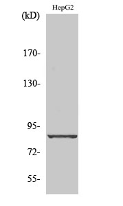
- Western Blot analysis of various cells using Cadherin-19 Polyclonal Antibody diluted at 1:1000
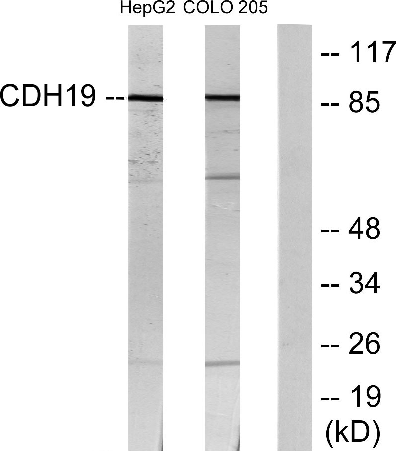
- Western blot analysis of lysates from HepG2 and COLO205 cells, using CDH19 Antibody. The lane on the right is blocked with the synthesized peptide.


