Cytokeratin (ABT154) mouse mAb
- 货号:YM6815
- 应用:IHC;ELISA
- 种属:Human;Mouse;Rat;
- 简介:
- >>Rap1 signaling pathway;>>Apelin signaling pathway;>>Hippo signaling pathway;>>Cell adhesion molecules;>>Adherens junction;>>Bacterial invasion of epithelial cells;>>Pathways in cancer;>>Endometrial cancer;>>Thyroid cancer;>>Melanoma;>>Bladder cancer;>>Gastric cancer
- 免疫原:
- Synthesized peptide derived from human Cytokeratin Pan AA range: 100-200
- 特异性:
- The antibody can recognize multiple human Cytokeratins, including CK10, 13, 14, 15, 16, 18, 19, and it can be used for immunohistochemical detection of tumors from monolayer and multilayered epitheliu
- 组成:
- PBS, 50% glycerol, 0.05% Proclin 300, 0.05%BSA
- 来源:
- Mouse, Monoclonal/IgG1, kappa
- 稀释:
- IHC 1:200-400. ELISA 1:500-5000
- 纯化工艺:
- The antibody was affinity-purified from ascites by affinity-chromatography using specific immunogen.
- 储存:
- -15°C to -25°C/1 year(Do not lower than -25°C)
- 细胞定位:
- Cytoplasmic, Membranous
- 组织表达:
- Non-neural epithelial tissues.
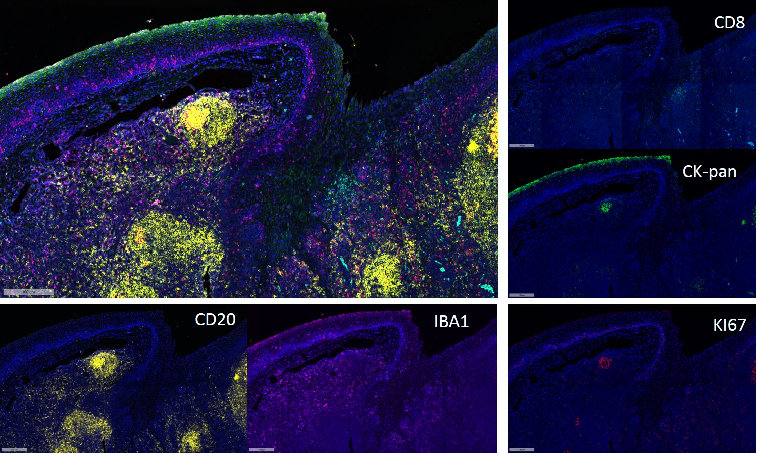
- Fluorescence multiplex immunohistochemical analysis of Human tonsil tissue (formalin-fixed paraffin-embedded section).
The immunostaining was performed by Pentuple-Fluorescence kit (RS0038, Immunoway). CK-pan mouse mAb(YM6815 Immunoway) green, Ki-67 rabbit mAb(YM7002 Immunoway) red, Iba 1 mouse mAb(YM4765
Immunoway) purple,CD8 a mouse mAb(YM4815
Immunoway) cyan, CD20 mouse mAb(YM4814 Immunoway) yellow,
The section was incubated in 5 rounds of staining; sequentially for Anti-antibodies; each using a separate fluorescent tyramide signal amplification system. EDTA based antigen retrieval (Immunoway YS0004, pH 9.0, 20 minutes) was used in between rounds of tyramide signal amplification to remove the antibody from the previous round, to avoid any cross-reactivity. DAPI (dark blue) was used as a nuclear counter stain.
Microscopy and pseudocoloring of individual dyes was performed using a Slideviewer Imaging System (Excilone).
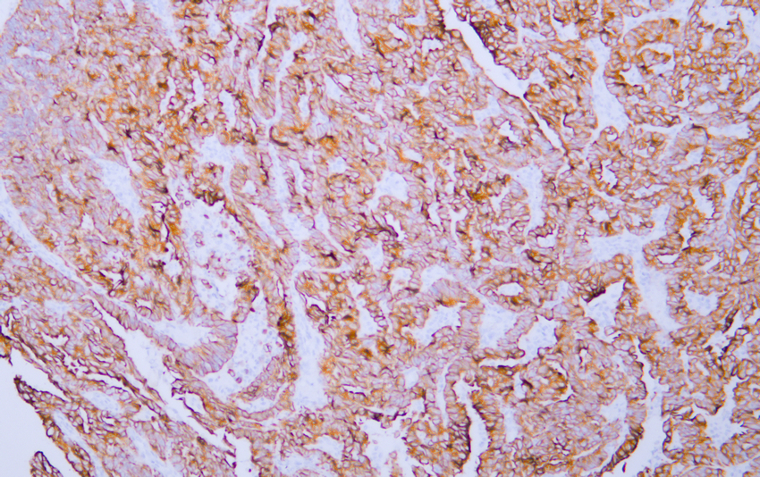
- Human endometrial adenocarcinoma tissue was stained with Anti-Cytokeratin (ABT154) Antibody

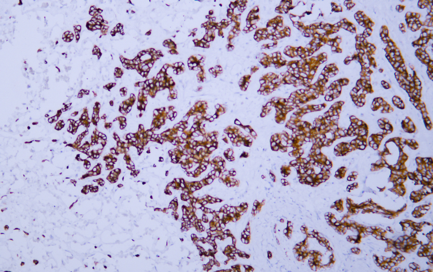
- Human esophageal squamous cell carcinoma tissue was stained with Anti-Cytokeratin (ABT154) Antibody
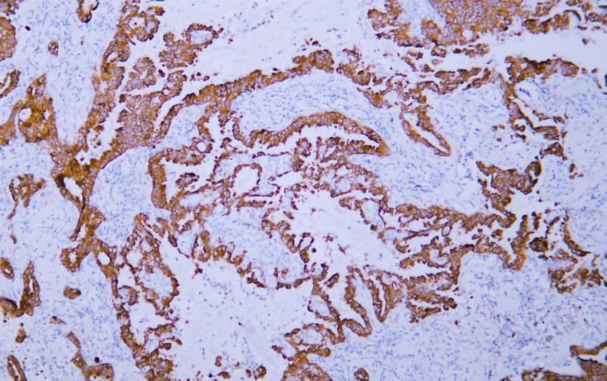
- Human hepatocellular carcinoma tissue was stained with Anti-Cytokeratin (ABT154) Antibody
- Human lung adenocarcinoma tissue was stained with Anti-Cytokeratin (ABT154) Antibody
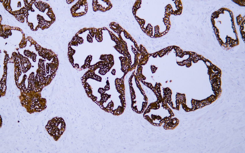
- Human prostate tissue was stained with Anti-Cytokeratin (ABT154) Antibody

- Fluorescence multiplex immunohistochemical analysis of normal human appendix tissue (formalin-fixed paraffin-embedded section).The section was incubated in 3 rounds of staining; in the order of CK PAN .( Catalog no:YM6815 1/200 dilution), PD-1.(Catalog no: YM6208 1/200 dilution), Caldesmon pan. (Catalog no:YM6826 1/200 dilution),each using a separate fluorescent tyramide signal amplification system : Treble-Fluorescence immunohistochemical mouse/rabbit kit Catalog NO: RS0035
(pH9.0)


