- 首页
- 公司介绍
- 热门促销
-
全部产品
-
试剂盒
- |
-
一抗
- |
-
二抗
- |
-
蛋白
- |
-
免疫组化试剂
- |
-
WB 试剂
- PonceauS Staining Solution
- PBST Washing Buffer, 10X
- 1.5M Tris-HCl Buffer, pH8.8
- 1M Tris-HCl Buffer, pH6.8
- 10% SDS Solution
- Prestained Protein Marker
- TBST Washing Buffer, 10X
- SDS PAGE Loading Buffer, 5X
- Stripping Buffered Solution
- Tris Buffer, pH7.4, 10X
- Total Protein Extraction Kit
- Running Buffer, 10X
- Transfer Buffer, 10X
- 30% Acr-Bis(29:1) Solution
- Tris电泳液速溶颗粒
- PBS(1X, premixed powder)
- TBS(1X, premixed powder)
- 快速封闭液
- 转膜液速溶颗粒
- Chemical reagents
- 公司新闻
- 营销网络
- 资源中心
- 联系我们
p21 (PTR2559) mouse mAb
- 货号:YM3802
- 应用:IHC;WB;IF;ELISA
- 种属:Human;Mouse;
- 基因名称:
- CDKN1A CAP20 CDKN1 CIP1 MDA6 PIC1 SDI1 WAF1
- 免疫原:
- Synthesized peptide derived from human p21 AA range: 1-100
- 特异性:
- This antibody detects endogenous levels of p21 protein.
- 组成:
- PBS, 50% glycerol, 0.05% Proclin 300, 0.05%BSA
- 来源:
- Mouse, Monoclonal/IgG1, kappa
- 稀释:
- IHC 1:200-1000. WB 1:500-2000. IF 1:100-500. ELISA 1:1000-5000
- 储存:
- -15°C to -25°C/1 year(Do not lower than -25°C)
- 其他名称:
- Cyclin-dependent kinase inhibitor 1 ;CDK-interacting protein 1;Melanoma differentiation-associated protein 6;MDA-6;p21;
- 功能:
- function:May be the important intermediate by which p53 mediates its role as an inhibitor of cellular proliferation in response to DNA damage. Binds to and inhibits cyclin-dependent kinase activity, preventing phosphorylation of critical cyclin-dependent kinase substrates and blocking cell cycle progression.,induction:By p53, mezerein (antileukemic compound) and interferon beta.,PTM:Phosphorylation of Thr-145 by Akt or of Ser-146 by PKC impairs binding to PCNA.,similarity:Belongs to the CDI family.,tissue specificity:Expressed in all adult human tissues, with 5-fold lower levels observed in the brain.,
- 细胞定位:
- Cytoplasmic, Nuclear
- 组织表达:
- Expressed in all adult tissues, with 5-fold lower levels observed in the brain.
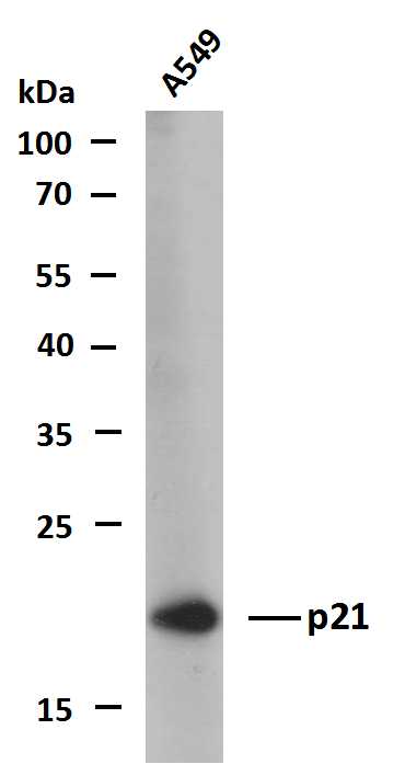
- Whole cell lysates of A549 were separated by 12% SDS-PAGE, and the membrane was blotted with anti-p21(PTR2559) antibody. The HRP-conjugated Goat anti-Mouse IgG(H + L) antibody was used to detect the antibody.
Lane 1: A549
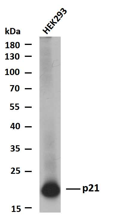
- Whole cell lysates of HEK293 were separated by 12% SDS-PAGE, and the membrane was blotted with anti-p21(PTR2559) antibody. The HRP-conjugated Goat anti-Mouse IgG(H + L) antibody was used to detect the antibody.
Lane 1: HEK293

- Whole cell lysates of MCF7 were separated by 12% SDS-PAGE, and the membrane was blotted with anti-p21(PTR2559) antibody. The HRP-conjugated Goat anti-Mouse IgG(H + L) antibody was used to detect the antibody.
Lane 1: MCF7


