NSE Monoclonal Antibody(13E2)
- 货号:YM3066
- 应用:WB;IF;IHC
- 种属:Human;Mouse;Rat
- 简介:
- >>Glycolysis / Gluconeogenesis;>>Metabolic pathways;>>Carbon metabolism;>>Biosynthesis of amino acids;>>RNA degradation;>>HIF-1 signaling pathway
- 免疫原:
- Synthetic Peptide of NSE
- 特异性:
- The antibody detects endogenous NSE proteins.
- 组成:
- PBS, pH 7.4, containing 0.5%BSA, 0.02% sodium azide as Preservative and 50% Glycerol.
- 稀释:
- WB 1:2000 IHC 1:200 IF 1:200
- 纯化工艺:
- The antibody was affinity-purified from mouse ascites by affinity-chromatography using specific immunogen.
- 储存:
- -15°C to -25°C/1 year(Do not lower than -25°C)
- 其他名称:
- ENO2;Gamma-enolase;2-phospho-D-glycerate hydro-lyase;Enolase 2;Neural enolase;Neuron-specific enolase;NSE
- 背景:
- enolase 2(ENO2) Homo sapiens This gene encodes one of the three enolase isoenzymes found in mammals. This isoenzyme, a homodimer, is found in mature neurons and cells of neuronal origin. A switch from alpha enolase to gamma enolase occurs in neural tissue during development in rats and primates. [provided by RefSeq, Jul 2008],
- 功能:
- catalytic activity:2-phospho-D-glycerate = phosphoenolpyruvate + H(2)O.,cofactor:Magnesium. Required for catalysis and for stabilizing the dimer.,developmental stage:During ontogenesis, there is a transition from the alpha/alpha homodimer to the alpha/beta heterodimer in striated muscle cells, and to the alpha/gamma heterodimer in nerve cells.,function:Has neurotrophic and neuroprotective properties on a broad spectrum of central nervous system (CNS) neurons. Binds, in a calcium-dependent manner, to cultured neocortical neurons and promotes cell survival.,induction:Levels of ENO2 increase dramatically in cardiovascular accidents, cerebral trauma, brain tumors and Creutzfeldt-Jacob disease.,pathway:Carbohydrate degradation; glycolysis; pyruvate from D-glyceraldehyde 3-phosphate: step 4/5.,similarity:Belongs to the enolase family.,subcellular location:Can translocate to the plasma membrane
- 细胞定位:
- Cytoplasm . Cell membrane . Can translocate to the plasma membrane in either the homodimeric (alpha/alpha) or heterodimeric (alpha/gamma) form. .
- 组织表达:
- The alpha/alpha homodimer is expressed in embryo and in most adult tissues. The alpha/beta heterodimer and the beta/beta homodimer are found in striated muscle, and the alpha/gamma heterodimer and the gamma/gamma homodimer in neurons.

- Immunofluorescence analysis of Hela cell. 1,Cdk2 Polyclonal Antibody(red) was diluted at 1:200(4° overnight). NSE Monoclonal Antibody(13E2)(green) was diluted at 1:200(4° overnight). 2, Goat Anti Rabbit Alexa Fluor 594 Catalog:RS3611 was diluted at 1:1000(room temperature, 50min). Goat Anti Mouse Alexa Fluor 488 Catalog:RS3208 was diluted at 1:1000(room temperature, 50min).
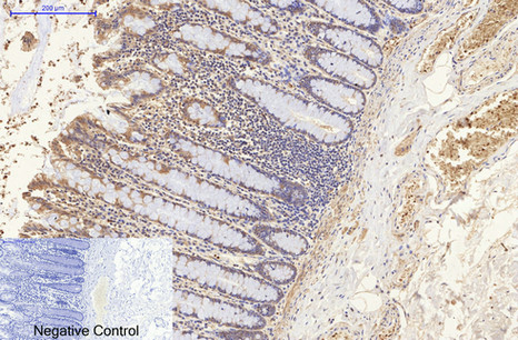
- Immunohistochemical analysis of paraffin-embedded Human-colon tissue. 1,NSE Monoclonal Antibody(13E2) was diluted at 1:200(4°C,overnight). 2, Sodium citrate pH 6.0 was used for antibody retrieval(>98°C,20min). 3,Secondary antibody was diluted at 1:200(room tempeRature, 30min). Negative control was used by secondary antibody only.
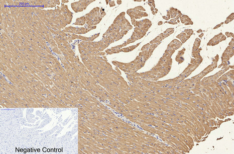
- Immunohistochemical analysis of paraffin-embedded Rat-heart tissue. 1,NSE Monoclonal Antibody(13E2) was diluted at 1:200(4°C,overnight). 2, Sodium citrate pH 6.0 was used for antibody retrieval(>98°C,20min). 3,Secondary antibody was diluted at 1:200(room tempeRature, 30min). Negative control was used by secondary antibody only.
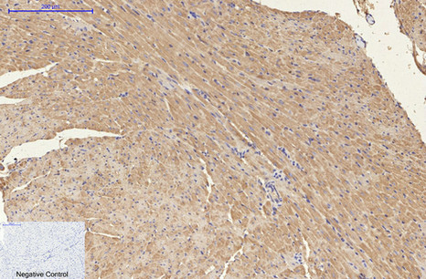
- Immunohistochemical analysis of paraffin-embedded Mouse-heart tissue. 1,NSE Monoclonal Antibody(13E2) was diluted at 1:200(4°C,overnight). 2, Sodium citrate pH 6.0 was used for antibody retrieval(>98°C,20min). 3,Secondary antibody was diluted at 1:200(room tempeRature, 30min). Negative control was used by secondary antibody only.
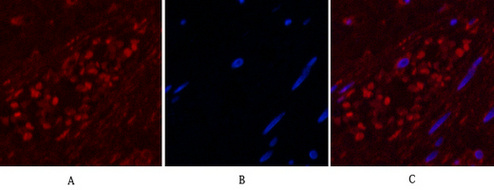
- Immunofluorescence analysis of Human-appendix tissue. 1,NSE Monoclonal Antibody(13E2)(red) was diluted at 1:200(4°C,overnight). 2, Cy3 labled Secondary antibody was diluted at 1:300(room temperature, 50min).3, Picture B: DAPI(blue) 10min. Picture A:Target. Picture B: DAPI. Picture C: merge of A+B
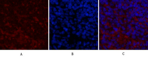
- Immunofluorescence analysis of Mouse-spleen tissue. 1,NSE Monoclonal Antibody(13E2)(red) was diluted at 1:200(4°C,overnight). 2, Cy3 labled Secondary antibody was diluted at 1:300(room temperature, 50min).3, Picture B: DAPI(blue) 10min. Picture A:Target. Picture B: DAPI. Picture C: merge of A+B
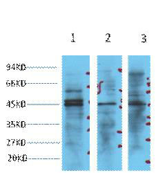
- Western blot analysis of 1) Hela, 2) Jurkat, 3) 293T cell lysates, diluted at 1:3000.
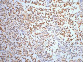
- IHC staining of Human small cell carcinoma of lung tissue, diluted at 1:200.
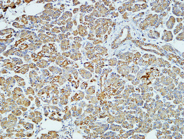
- Immunohistochemical analysis of paraffin-embedded Human pancreas. 1, Antibody was diluted at 1:100(4° overnight). 2, High-pressure and temperature EDTA, pH8.0 was used for antigen retrieval. 3,Secondary antibody was diluted at 1:200(room temperature, 30min).
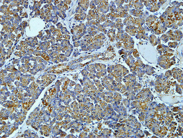
- Immunohistochemical analysis of paraffin-embedded Human pancreas. 1, Antibody was diluted at 1:400(4° overnight). 2, High-pressure and temperature EDTA, pH8.0 was used for antigen retrieval. 3,Secondary antibody was diluted at 1:200(room temperature, 30min).
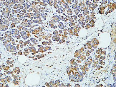
- Immunohistochemical analysis of paraffin-embedded Human pancreas. 1, Antibody was diluted at 1:400(4° overnight). 2, High-pressure and temperature EDTA, pH8.0 was used for antigen retrieval. 3,Secondary antibody was diluted at 1:200(room temperature, 30min).
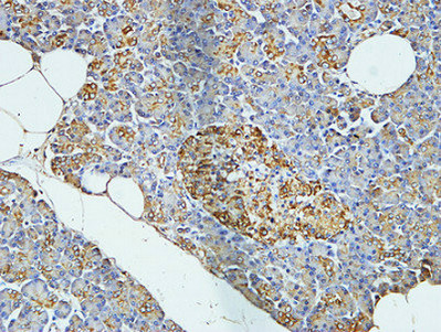
- Immunohistochemical analysis of paraffin-embedded Human pancreas. 1, Antibody was diluted at 1:400(4° overnight). 2, High-pressure and temperature EDTA, pH8.0 was used for antigen retrieval. 3,Secondary antibody was diluted at 1:200(room temperature, 30min).


