Kif 7 Monoclonal Antibody(3F8)
- 货号:YM3063
- 应用:IHC;IF
- 种属:Human;Mouse;Rat
- 简介:
- >>Hedgehog signaling pathway;>>Pathways in cancer;>>Basal cell carcinoma
- 蛋白名称:
- Kinesin-like protein KIF7
- 免疫原:
- Synthetic Peptide of Kif 7
- 特异性:
- The antibody detects endogenous Kif 7 proteins.
- 组成:
- PBS, pH 7.4, containing 0.5%BSA, 0.02% sodium azide as Preservative and 50% Glycerol.
- 稀释:
- IHC 1:50-200. IF 1:50-200
- 纯化工艺:
- The antibody was affinity-purified from mouse ascites by affinity-chromatography using specific immunogen.
- 储存:
- -15°C to -25°C/1 year(Do not lower than -25°C)
- 其他名称:
- Kinesin-like protein KIF7
- 背景:
- This gene encodes a cilia-associated protein belonging to the kinesin family. This protein plays a role in the sonic hedgehog (SHH) signaling pathway through the regulation of GLI transcription factors. It functions as a negative regulator of the SHH pathway by preventing inappropriate activation of GLI2 in the absence of ligand, and as a positive regulator by preventing the processing of GLI3 into its repressor form. Mutations in this gene have been associated with various ciliopathies. [provided by RefSeq, Oct 2011],
- 功能:
- similarity:Belongs to the kinesin-like protein family. KIF27 subfamily.,similarity:Contains 1 kinesin-motor domain.,tissue specificity:Embryonic stem cells, melanotic melanoma and Jurkat T-cells.,
- 细胞定位:
- Cell projection, cilium . Cytoplasm, cytoskeleton, cilium basal body . Localizes to the cilium tip.
- 组织表达:
- Embryonic stem cells, melanotic melanoma and Jurkat T-cells. Expressed in heart, lung, liver, kidney, testis, retina, placenta, pancreas, colon, small intestin, prostate and thymus.
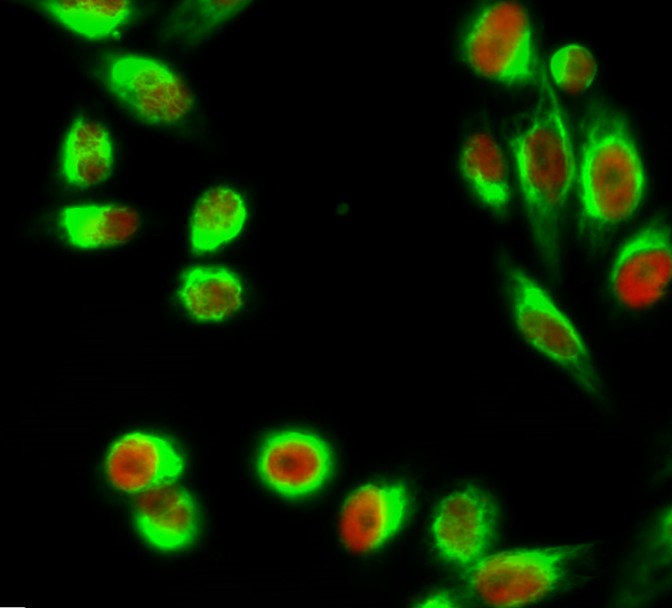
- Immunofluorescence analysis of Hela cell. 1,C/EBP β Polyclonal Antibody(red) was diluted at 1:200(4° overnight). Kif 7 Monoclonal Antibody(3F8)(green) was diluted at 1:200(4° overnight). 2, Goat Anti Rabbit Alexa Fluor 594 Catalog:RS3611 was diluted at 1:1000(room temperature, 50min). Goat Anti Mouse Alexa Fluor 488 Catalog:RS3208 was diluted at 1:1000(room temperature, 50min).
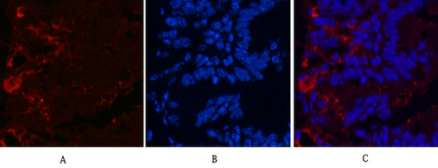
- Immunofluorescence analysis of Mouse-colon tissue. 1,Kif 7 Monoclonal Antibody(3F8)(red) was diluted at 1:200(4°C,overnight). 2, Cy3 labled Secondary antibody was diluted at 1:300(room temperature, 50min).3, Picture B: DAPI(blue) 10min. Picture A:Target. Picture B: DAPI. Picture C: merge of A+B
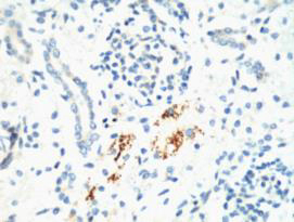
- IHC staining of Mouse Kidney tissue, diluted at 1:200.
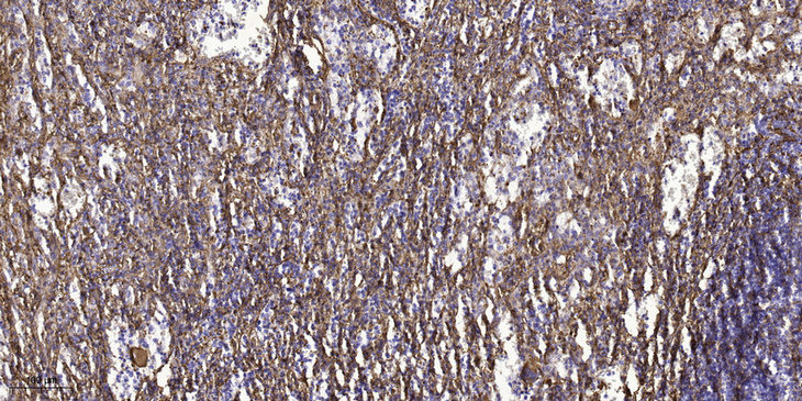
- Immunohistochemical analysis of paraffin-embedded human spleen tissue. 1,primary Antibody was diluted at 1:200(4° overnight). 2, Sodium citrate pH 6.0 was used for antigen retrieval(>98°C,20min). 3,Secondary antibody was diluted at 1:200


