- 首页
- 公司介绍
- 热门促销
-
全部产品
-
试剂盒
- |
-
一抗
- |
-
二抗
- |
-
蛋白
- |
-
免疫组化试剂
- |
-
WB 试剂
- PonceauS Staining Solution
- PBST Washing Buffer, 10X
- 1.5M Tris-HCl Buffer, pH8.8
- 1M Tris-HCl Buffer, pH6.8
- 10% SDS Solution
- Prestained Protein Marker
- TBST Washing Buffer, 10X
- SDS PAGE Loading Buffer, 5X
- Stripping Buffered Solution
- Tris Buffer, pH7.4, 10X
- Total Protein Extraction Kit
- Running Buffer, 10X
- Transfer Buffer, 10X
- 30% Acr-Bis(29:1) Solution
- Tris电泳液速溶颗粒
- PBS(1X, premixed powder)
- TBS(1X, premixed powder)
- 快速封闭液
- 转膜液速溶颗粒
- Chemical reagents
- 公司新闻
- 营销网络
- 资源中心
- 联系我们
Hck Monoclonal Antibody
- 货号:YM0326
- 应用:WB;IHC;IF;ELISA
- 种属:Human
- 简介:
- >>Chemokine signaling pathway;>>Fc gamma R-mediated phagocytosis;>>Kaposi sarcoma-associated herpesvirus infection
- 蛋白名称:
- Tyrosine-protein kinase HCK
- 免疫原:
- Purified recombinant fragment of Hck expressed in E. Coli.
- 特异性:
- Hck Monoclonal Antibody detects endogenous levels of Hck protein.
- 组成:
- Liquid in PBS containing 50% glycerol, 0.5% BSA and 0.02% sodium azide.
- 稀释:
- WB 1:500 - 1:2000. IHC 1:200 - 1:1000. ELISA: 1:10000.. IF 1:50-200
- 纯化工艺:
- Affinity purification
- 储存:
- -15°C to -25°C/1 year(Do not lower than -25°C)
- 其他名称:
- HCK;Tyrosine-protein kinase HCK;Hematopoietic cell kinase;Hemopoietic cell kinase;p59-HCK/p60-HCK;p59Hck;p61Hck
- 背景:
- The protein encoded by this gene is a member of the Src family of tyrosine kinases. This protein is primarily hemopoietic, particularly in cells of the myeloid and B-lymphoid lineages. It may help couple the Fc receptor to the activation of the respiratory burst. In addition, it may play a role in neutrophil migration and in the degranulation of neutrophils. Multiple isoforms with different subcellular distributions are produced due to both alternative splicing and the use of alternative translation initiation codons, including a non-AUG (CUG) codon. [provided by RefSeq, Feb 2010],
- 功能:
- catalytic activity:ATP + a [protein]-L-tyrosine = ADP + a [protein]-L-tyrosine phosphate.,domain:The SH3 domain mediates binding to HIV-1 Nef.,function:May serve as part of a signaling pathway coupling the Fc receptor to the activation of the respiratory burst. May also contribute to neutrophil migration and may regulate the degranulation process of neutrophils.,PTM:Isoform p59-HCK contains a N-myristoyl glycine at position 3 (By similarity). Isoform p59-HCK contains a S-palmitoyl cysteine at position 3.,similarity:Belongs to the protein kinase superfamily. Tyr protein kinase family. SRC subfamily.,similarity:Contains 1 protein kinase domain.,similarity:Contains 1 SH2 domain.,similarity:Contains 1 SH3 domain.,subunit:May interact (via SH3 domain) with HIV-1 Nef and Vif. This interaction would stimulates its tyrosine-kinase activity. Interacts (via SH3 domain) with HEV ORF3 protein.,tissu
- 细胞定位:
- [Isoform 1]: Lysosome. Membrane; Lipid-anchor. Cell projection, podosome membrane; Lipid-anchor. Cytoplasm, cytosol. Associated with specialized secretory lysosomes called azurophil granules. At least half of this isoform is found in the cytoplasm, some of this fraction is myristoylated.; [Isoform 2]: Cell membrane ; Lipid-anchor . Membrane, caveola ; Lipid-anchor . Cell junction, focal adhesion . Cytoplasm, cytoskeleton . Golgi apparatus . Cytoplasmic vesicle . Lysosome . Nucleus . 20% of this isoform is associated with caveolae. Localization at the cell membrane and at caveolae requires palmitoylation at Cys-3. Colocalizes with the actin cytoskeleton at focal adhesions.; Cytoplasmic vesicle, secretory vesicle. Cytoplasm, cytosol.
- 组织表达:
- Detected in monocytes and neutrophils (at protein level). Expressed predominantly in cells of the myeloid and B-lymphoid lineages. Highly expressed in granulocytes. Detected in tonsil.
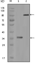
- Western Blot analysis using Hck Monoclonal Antibody against truncated HCK recombinant protein (1) and full-length HCK-GFP transfected CHO-K1 cell lysate (2).
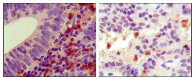
- Immunohistochemistry analysis of paraffin-embedded human colon cancer (left) and ancreas cancer (right), showing cytoplasmic localization with DAB staining using Hck Monoclonal Antibody.


