- 首页
- 公司介绍
- 热门促销
-
全部产品
-
试剂盒
- |
-
一抗
- |
-
二抗
- |
-
蛋白
- |
-
免疫组化试剂
- |
-
WB 试剂
- PonceauS Staining Solution
- PBST Washing Buffer, 10X
- 1.5M Tris-HCl Buffer, pH8.8
- 1M Tris-HCl Buffer, pH6.8
- 10% SDS Solution
- Prestained Protein Marker
- TBST Washing Buffer, 10X
- SDS PAGE Loading Buffer, 5X
- Stripping Buffered Solution
- Tris Buffer, pH7.4, 10X
- Total Protein Extraction Kit
- Running Buffer, 10X
- Transfer Buffer, 10X
- 30% Acr-Bis(29:1) Solution
- Tris电泳液速溶颗粒
- PBS(1X, premixed powder)
- TBS(1X, premixed powder)
- 快速封闭液
- 转膜液速溶颗粒
- Chemical reagents
- 公司新闻
- 营销网络
- 资源中心
- 联系我们
CD8 a (ABT304) IHC kit
- 货号:IHCM6938
- 应用:IHC
- 种属:Human;
- 简介:
- >>Cell adhesion molecules;>>Antigen processing and presentation;>>Hematopoietic cell lineage;>>T cell receptor signaling pathway;>>Yersinia infection;>>Primary immunodeficiency
- 蛋白名称:
- alpha polypeptide (p32);CD_antigen=CD8a;CD8;CD8 antigen alpha polypeptide;CD8 antigen alpha polypeptide (p32);CD8 antigen, alpha polypeptide (p32);CD8a;CD8A antigen;CD8A molecule;CD8A_HUMAN;Leu2;Leu2
- 免疫原:
- Synthesized peptide derived from human CD8 AA range: 100-235
- 特异性:
- The antibody can specifically recognize human CD8 protein, including two typies of dimer: αβ heterodimer or αα homodimer.
- 来源:
- Mouse, Monoclonal/IgG2b, kappa
- 纯化工艺:
- The antibody was affinity-purified from ascites by affinity-chromatography using specific immunogen.
- 其他名称:
- alpha polypeptide (p32);CD_antigen=CD8a;CD8;CD8 antigen alpha polypeptide;CD8 antigen alpha polypeptide (p32);CD8 antigen, alpha polypeptide (p32);CD8a;CD8A antigen;CD8A molecule;CD8A_HUMAN;Leu2;Leu2 T lymphocyte antigen;Ly 2;Ly 35;Ly B;Ly2;Ly3;Ly35;LyB;Lyt 2.1 lymphocyte differentiation antigen (AA at 100);LYT3;MAL;OKT8 T cell antigen;OTTHUMP00000160760;OTTHUMP00000160764;OTTHUMP00000203528;OTTHUMP00000203721;p32;T cell antigen Leu2;T cell co receptor;T lymphocyte differentiation antigen T8/Leu 2;T-cell surface glycoprotein CD8 alpha chain;T-cell surface glycoprotein Lyt 2;T-lymphocyte differentiation antigen T8/Leu-2;T8 T cell antigen;T8/Leu-2 T-lymphocyte differentiation antigen
- 背景:
- The CD8 antigen is a cell surface glycoprotein found on most cytotoxic T lymphocytes that mediates efficient cell-cell interactions within the immune system. The CD8 antigen acts as a coreceptor with the T-cell receptor on the T lymphocyte to recognize antigens displayed by an antigen presenting cell in the context of class I MHC molecules. The coreceptor functions as either a homodimer composed of two alpha chains or as a heterodimer composed of one alpha and one beta chain. Both alpha and beta chains share significant homology to immunoglobulin variable light chains. This gene encodes the CD8 alpha chain. Multiple transcript variants encoding different isoforms have been found for this gene. [provided by RefSeq, Nov 2011],
- 功能:
- disease:Defects in CD8A are a cause of familial CD8 deficiency (CD8 deficiency) [MIM:608957]. Familial CD8 deficiency is a novel autosomal recessive immunologic defect characterized by absence of CD8+ cells, leading to recurrent bacterial infections.,function:Identifies cytotoxic/suppressor T-cells that interact with MHC class I bearing targets. CD8 is thought to play a role in the process of T-cell mediated killing. CD8 alpha chains binds to class I MHC molecules alpha-3 domains.,online information:CD8 entry,online information:CD8A mutation db,PTM:All of the five most carboxyl-terminal cysteines form inter-chain disulfide bonds in dimers and higher multimers, while the four N-terminal cysteines do not.,similarity:Contains 1 Ig-like V-type (immunoglobulin-like) domain.,subunit:In general heterodimer of an alpha and a beta chain linked by two disulfide bonds. Can also form homodimers. Sho

- Human appendix tissue was stained with Anti-CD8 (ABT304) Antibody

- Human burkitt lymphoma tissue was stained with Anti-CD8 (ABT304) Antibody
.jpg)
- Human lymphoma tissue was stained with Anti-CD8 (ABT304) Antibody
.jpg)
- Human lymphoma tissue was stained with Anti-CD8 (ABT304) Antibody

- Human tonsil tissue was stained with Anti-CD8 (ABT304) Antibody

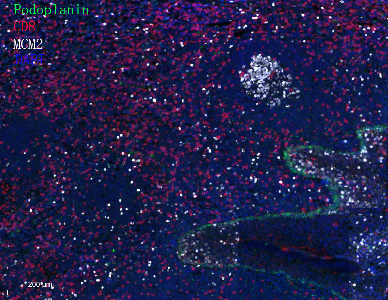
- Fluorescence multiplex immunohistochemical analysis of Human tonsil tissue (formalin-fixed paraffin-embedded section).
Merged staining of Anti-Podoplanin (YM6994), Anti-CD8 (YM6938), Anti-MCM2 (YM6077).
The immunostaining was performed on a Leica Biosystems BOND® MAX instrument with an Sextuple-Fluorescence kit (RS0039, Immunoway).
The section was incubated in 3 rounds of staining; sequentially for Anti-Podoplanin (YM6994 1:200), Anti-CD8 (YM6938 1:200), Anti-MCM2 (YM6077 1:200).; each using a separate fluorescent tyramide signal amplification system. EDTA based antigen retrieval (Leica Biosystems BOND® Epitope Retrieval Solution 2, pH 9.0, 20 minutes) was used in between rounds of tyramide signal amplification to remove the antibody from the previous round, to avoid any cross-reactivity. DAPI (dark blue) was used as a nuclear counter stain.
Microscopy and pseudocoloring of individual dyes was performed using a Slideviewer Imaging System (3D histech).

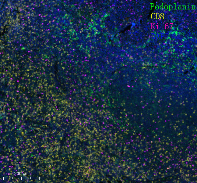
- Fluorescence multiplex immunohistochemical analysis of Human tonsil tissue (formalin-fixed paraffin-embedded section).
Merged staining of Anti-Podoplanin (YM6994), Anti-CD8 (YM6938), Anti-Ki-67 (YM6812).
The immunostaining was performed on a Leica Biosystems BOND® MAX instrument with an Sextuple-Fluorescence kit (RS0039, Immunoway).
The section was incubated in 3 rounds of staining; sequentially for Anti-Podoplanin (YM6994 1:200), Anti-CD8 (YM6938 1:200), Anti-Ki67 (YM6812 1:200).; each using a separate fluorescent tyramide signal amplification system. EDTA based antigen retrieval (Leica Biosystems BOND® Epitope Retrieval Solution 2, pH 9.0, 20 minutes) was used in between rounds of tyramide signal amplification to remove the antibody from the previous round, to avoid any cross-reactivity. DAPI (dark blue) was used as a nuclear counter stain.
Microscopy and pseudocoloring of individual dyes was performed using a Slideviewer Imaging System (3D histech).

- Fluorescence multiplex immunohistochemical analysis of Human tonsil tissue (formalin-fixed paraffin-embedded section).
Merged staining of Anti-Podoplanin (YM6994), Anti-CD8 (YM6938), Anti-MCM2 (YM6077).
The immunostaining was performed on a Leica Biosystems BOND® MAX instrument with an Sextuple-Fluorescence kit (RS0039, Immunoway).
The section was incubated in 3 rounds of staining; sequentially for Anti-Podoplanin (YM6994 1:200), Anti-CD8 (YM6938 1:200), Anti-MCM2 (YM6077 1:200).; each using a separate fluorescent tyramide signal amplification system. EDTA based antigen retrieval (Leica Biosystems BOND® Epitope Retrieval Solution 2, pH 9.0, 20 minutes) was used in between rounds of tyramide signal amplification to remove the antibody from the previous round, to avoid any cross-reactivity. DAPI (dark blue) was used as a nuclear counter stain.
Microscopy and pseudocoloring of individual dyes was performed using a Slideviewer Imaging System (3D histech).


.jpg)
.jpg)


