V-ATPase H Polyclonal Antibody
- Catalog No.:YT4861
- Applications:WB;IHC;IF;ELISA
- Reactivity:Human;Mouse
- Target:
- V-ATPase H
- Fields:
- >>Oxidative phosphorylation;>>Metabolic pathways;>>Lysosome;>>Phagosome;>>mTOR signaling pathway;>>Synaptic vesicle cycle;>>Vibrio cholerae infection;>>Epithelial cell signaling in Helicobacter pylori infection;>>Tuberculosis;>>Human papillomavirus infection;>>Rheumatoid arthritis
- Gene Name:
- ATP6V1H
- Protein Name:
- V-type proton ATPase subunit H
- Human Gene Id:
- 51606
- Human Swiss Prot No:
- Q9UI12
- Mouse Gene Id:
- 108664
- Mouse Swiss Prot No:
- Q8BVE3
- Immunogen:
- The antiserum was produced against synthesized peptide derived from human ATP6V1H. AA range:341-390
- Specificity:
- V-ATPase H Polyclonal Antibody detects endogenous levels of V-ATPase H protein.
- Formulation:
- Liquid in PBS containing 50% glycerol, 0.5% BSA and 0.02% sodium azide.
- Source:
- Polyclonal, Rabbit,IgG
- Dilution:
- WB 1:500 - 1:2000. IHC 1:100 - 1:300. ELISA: 1:20000.. IF 1:50-200
- Purification:
- The antibody was affinity-purified from rabbit antiserum by affinity-chromatography using epitope-specific immunogen.
- Concentration:
- 1 mg/ml
- Storage Stability:
- -15°C to -25°C/1 year(Do not lower than -25°C)
- Other Name:
- ATP6V1H;CGI-11;V-type proton ATPase subunit H;V-ATPase subunit H;Nef-binding protein 1;NBP1;Protein VMA13 homolog;V-ATPase 50/57 kDa subunits;Vacuolar proton pump subunit H;Vacuolar proton pump subunit SFD
- Observed Band(KD):
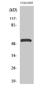
- 55kD
- Background:
- This gene encodes a component of vacuolar ATPase (V-ATPase), a multisubunit enzyme that mediates acidification of intracellular organelles. V-ATPase-dependent organelle acidification is necessary for multiple processes including protein sorting, zymogen activation, receptor-mediated endocytosis, and synaptic vesicle proton gradient generation. The encoded protein is the regulatory H subunit of the V1 domain of V-ATPase, which is required for catalysis of ATP but not the assembly of V-ATPase. Decreased expression of this gene may play a role in the development of type 2 diabetes. Alternatively spliced transcript variants encoding multiple isoforms have been observed for this gene. [provided by RefSeq, May 2012],
- Function:
- function:Subunit of the peripheral V1 complex of vacuolar ATPase. Subunit H activates the ATPase activity of the enzyme and couples ATPase activity to proton flow. Vacuolar ATPase is responsible for acidifying a variety of intracellular compartments in eukaryotic cells, thus providing most of the energy required for transport processes in the vacuolar system (By similarity). Involved in the endocytosis mediated by clathrin-coated pits, required for the formation of endosomes.,similarity:Belongs to the V-ATPase H subunit family.,subunit:V-ATPase is an heteromultimeric enzyme composed of a peripheral catalytic V1 complex (components A to H) attached to an integral membrane V0 proton pore complex (components: a, c, c', c'' and d). Interacts with HIV-1 Nef protein and AP2M1.,tissue specificity:Widely expressed.,
- Subcellular Location:
- Cytoplasmic vesicle, clathrin-coated vesicle membrane ; Peripheral membrane protein .
- Expression:
- Widely expressed.
- June 19-2018
- WESTERN IMMUNOBLOTTING PROTOCOL
- June 19-2018
- IMMUNOHISTOCHEMISTRY-PARAFFIN PROTOCOL
- June 19-2018
- IMMUNOFLUORESCENCE PROTOCOL
- September 08-2020
- FLOW-CYTOMEYRT-PROTOCOL
- May 20-2022
- Cell-Based ELISA│解您多样本WB检测之困扰
- July 13-2018
- CELL-BASED-ELISA-PROTOCOL-FOR-ACETYL-PROTEIN
- July 13-2018
- CELL-BASED-ELISA-PROTOCOL-FOR-PHOSPHO-PROTEIN
- July 13-2018
- Antibody-FAQs
- Products Images

- Western Blot analysis of various cells using V-ATPase H Polyclonal Antibody. Secondary antibody(catalog#:RS0002) was diluted at 1:20000

- Immunohistochemistry analysis of paraffin-embedded human brain tissue, using ATP6V1H Antibody. The picture on the right is blocked with the synthesized peptide.


