Pim-2 Polyclonal Antibody
- Catalog No.:YT3729
- Applications:IHC;IF;ELISA
- Reactivity:Human;Rat;Mouse;
- Target:
- Pim-2
- Fields:
- >>Pathways in cancer;>>Acute myeloid leukemia
- Gene Name:
- PIM2
- Protein Name:
- Serine/threonine-protein kinase pim-2
- Human Gene Id:
- 11040
- Human Swiss Prot No:
- Q9P1W9
- Mouse Swiss Prot No:
- Q62070
- Immunogen:
- Synthesized peptide derived from the C-terminal region of human Pim-2.
- Specificity:
- Pim-2 Polyclonal Antibody detects endogenous levels of Pim-2 protein.
- Formulation:
- Liquid in PBS containing 50% glycerol, 0.5% BSA and 0.02% sodium azide.
- Source:
- Polyclonal, Rabbit,IgG
- Dilution:
- IHC 1:100 - 1:300. ELISA: 1:10000.. IF 1:50-200
- Purification:
- The antibody was affinity-purified from rabbit antiserum by affinity-chromatography using epitope-specific immunogen.
- Concentration:
- 1 mg/ml
- Storage Stability:
- -15°C to -25°C/1 year(Do not lower than -25°C)
- Other Name:
- PIM2;Serine/threonine-protein kinase pim-2;Pim-2h
- Molecular Weight(Da):
- 34kD
- Background:
- This gene encodes a protooncogene that acts as a serine/threonine protein kinase. Studies determined the encoded protein functions to prevent apoptosis and to promote cell survival.[provided by RefSeq, Nov 2009],
- Function:
- catalytic activity:ATP + a protein = ADP + a phosphoprotein.,similarity:Belongs to the protein kinase superfamily.,similarity:Belongs to the protein kinase superfamily. CAMK Ser/Thr protein kinase family. PIM subfamily.,similarity:Contains 1 protein kinase domain.,tissue specificity:Highly expressed in hematopoietic tissues, in leukemic and lymphoma cell lines, testis, small intestine, colon and colorectal adenocarcinoma cells.,
- Subcellular Location:
- cytoplasm,
- Expression:
- Highly expressed in hematopoietic tissues, in leukemic and lymphoma cell lines, testis, small intestine, colon and colorectal adenocarcinoma cells. Weakly expressed in normal liver, but highly expressed in hepatocellular carcinoma tissues.
- June 19-2018
- WESTERN IMMUNOBLOTTING PROTOCOL
- June 19-2018
- IMMUNOHISTOCHEMISTRY-PARAFFIN PROTOCOL
- June 19-2018
- IMMUNOFLUORESCENCE PROTOCOL
- September 08-2020
- FLOW-CYTOMEYRT-PROTOCOL
- May 20-2022
- Cell-Based ELISA│解您多样本WB检测之困扰
- July 13-2018
- CELL-BASED-ELISA-PROTOCOL-FOR-ACETYL-PROTEIN
- July 13-2018
- CELL-BASED-ELISA-PROTOCOL-FOR-PHOSPHO-PROTEIN
- July 13-2018
- Antibody-FAQs
- Products Images
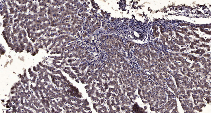
- Immunohistochemical analysis of paraffin-embedded human liver cancer. 1, Antibody was diluted at 1:200(4° overnight). 2, Tris-EDTA,pH9.0 was used for antigen retrieval. 3,Secondary antibody was diluted at 1:200(room temperature, 45min).


