Neuroglycan C Polyclonal Antibody
- Catalog No.:YT3066
- Applications:WB;IHC
- Reactivity:Human;Mouse;Rat
- Target:
- Neuroglycan C
- Gene Name:
- CSPG5
- Protein Name:
- Chondroitin sulfate proteoglycan 5
- Human Gene Id:
- 10675
- Human Swiss Prot No:
- O95196
- Mouse Gene Id:
- 29873
- Mouse Swiss Prot No:
- Q71M36
- Rat Gene Id:
- 50568
- Rat Swiss Prot No:
- Q9ERQ6
- Immunogen:
- The antiserum was produced against synthesized peptide derived from human CSPG5. AA range:211-260
- Specificity:
- Neuroglycan C Polyclonal Antibody detects endogenous levels of Neuroglycan C protein.
- Formulation:
- Liquid in PBS containing 50% glycerol, 0.5% BSA and 0.02% sodium azide.
- Source:
- Polyclonal, Rabbit,IgG
- Dilution:
- WB 1:500-2000;IHC 1:50-300
- Purification:
- The antibody was affinity-purified from rabbit antiserum by affinity-chromatography using epitope-specific immunogen.
- Concentration:
- 1 mg/ml
- Storage Stability:
- -15°C to -25°C/1 year(Do not lower than -25°C)
- Other Name:
- CSPG5;CALEB;NGC;Chondroitin sulfate proteoglycan 5;Acidic leucine-rich EGF-like domain-containing brain protein;Neuroglycan C
- Observed Band(KD):
- 60kD
- Background:
- The protein encoded by this gene is a proteoglycan that may function as a neural growth and differentiation factor. Several transcript variants encoding different isoforms have been found for this gene. [provided by RefSeq, May 2011],
- Function:
- developmental stage:Expressed in brain of 3 months, 5 and 10-year-old individuals.,function:May function as a growth and differentiation factor involved in neuritogenesis. May induce ERBB3 activation.,miscellaneous:Different forms of various molecular weight have been observed. Such forms are possibly due to different levels of glycosylation, phosphorylation and/or protein cleavage.,PTM:N-glycosylated.,PTM:O-glycosylated; contains chondroitin sulfate glycans. Part-time proteoglycan, expressed in part as a proteoglycan exhibiting chondroitin sulfate glycans and in part as a non-proteoglycan form. The relative amount of both forms depends on tissues and tissues maturation.,PTM:Phosphorylated; in intracellular and extracellular parts.,similarity:Contains 1 EGF-like domain.,subcellular location:In neurons, localizes to synaptic junctions (By similarity). Also detected in the endoplasmic reti
- Subcellular Location:
- Cell membrane ; Single-pass type I membrane protein . Cell junction, synapse, synaptic cell membrane ; Single-pass type I membrane protein . Endoplasmic reticulum membrane ; Single-pass type I membrane protein . Golgi apparatus membrane ; Single-pass type I membrane protein . Cell surface . In neurons, localizes to synaptic junctions. Also detected in the endoplasmic reticulum and the Golgi. Partially enriched in lipid rafts. .
- Expression:
- Restricted to brain (at protein level).
- June 19-2018
- WESTERN IMMUNOBLOTTING PROTOCOL
- June 19-2018
- IMMUNOHISTOCHEMISTRY-PARAFFIN PROTOCOL
- June 19-2018
- IMMUNOFLUORESCENCE PROTOCOL
- September 08-2020
- FLOW-CYTOMEYRT-PROTOCOL
- May 20-2022
- Cell-Based ELISA│解您多样本WB检测之困扰
- July 13-2018
- CELL-BASED-ELISA-PROTOCOL-FOR-ACETYL-PROTEIN
- July 13-2018
- CELL-BASED-ELISA-PROTOCOL-FOR-PHOSPHO-PROTEIN
- July 13-2018
- Antibody-FAQs
- Products Images

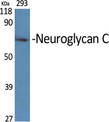
- Western Blot analysis of various cells using Neuroglycan C Polyclonal Antibody
.jpg)
- Western Blot analysis of HeLa cells using Neuroglycan C Polyclonal Antibody

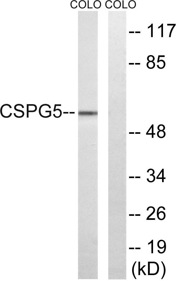
- Western blot analysis of lysates from COLO and HeLa cells, using CSPG5 Antibody. The lane on the right is blocked with the synthesized peptide.

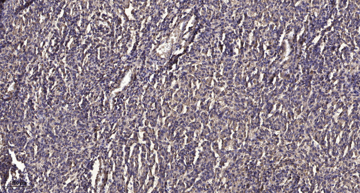
- Immunohistochemical analysis of paraffin-embedded human meningioma. 1, Antibody was diluted at 1:200(4° overnight). 2, Tris-EDTA,pH9.0 was used for antigen retrieval. 3,Secondary antibody was diluted at 1:200(room temperature, 45min).


