TGFβ RI (phospho Ser165) Polyclonal Antibody
- Catalog No.:YP1191
- Applications:WB;IHC;IF;ELISA
- Reactivity:Human;Mouse;Rat
- Target:
- TGF β Receptor I
- Fields:
- >>MAPK signaling pathway;>>Cytokine-cytokine receptor interaction;>>FoxO signaling pathway;>>Endocytosis;>>Cellular senescence;>>TGF-beta signaling pathway;>>Apelin signaling pathway;>>Osteoclast differentiation;>>Hippo signaling pathway;>>Adherens junction;>>Th17 cell differentiation;>>Relaxin signaling pathway;>>AGE-RAGE signaling pathway in diabetic complications;>>Chagas disease;>>Hepatitis B;>>Human T-cell leukemia virus 1 infection;>>Pathways in cancer;>>Colorectal cancer;>>Pancreatic cancer;>>Chronic myeloid leukemia;>>Hepatocellular carcinoma;>>Gastric cancer;>>Diabetic cardiomyopathy
- Gene Name:
- TGFBR1
- Protein Name:
- TGF-beta receptor type-1
- Human Gene Id:
- 7046
- Human Swiss Prot No:
- P36897
- Mouse Gene Id:
- 21812
- Mouse Swiss Prot No:
- Q64729
- Rat Swiss Prot No:
- P80204
- Immunogen:
- The antiserum was produced against synthesized peptide derived from human TGF beta Receptor I around the phosphorylation site of Ser165. AA range:131-180
- Specificity:
- Phospho-TGFβ RI (S165) Polyclonal Antibody detects endogenous levels of TGFβ RI protein only when phosphorylated at S165.
- Formulation:
- Liquid in PBS containing 50% glycerol, 0.5% BSA and 0.02% sodium azide.
- Source:
- Polyclonal, Rabbit,IgG
- Dilution:
- WB 1:500-2000, IF 1:50-300, IHC 1:50-300
- Purification:
- The antibody was affinity-purified from rabbit antiserum by affinity-chromatography using epitope-specific immunogen.
- Concentration:
- 1 mg/ml
- Storage Stability:
- -15°C to -25°C/1 year(Do not lower than -25°C)
- Other Name:
- TGFBR1;ALK5;SKR4;TGF-beta receptor type-1;TGFR-1;Activin A receptor type II-like protein kinase of 53kD;Activin receptor-like kinase 5;ALK-5;ALK5;Serine/threonine-protein kinase receptor R4;SKR4;TGF-beta type I receptor;Transfor
- Molecular Weight(Da):
- 56kD
- Background:
- The protein encoded by this gene forms a heteromeric complex with type II TGF-beta receptors when bound to TGF-beta, transducing the TGF-beta signal from the cell surface to the cytoplasm. The encoded protein is a serine/threonine protein kinase. Mutations in this gene have been associated with Loeys-Dietz aortic aneurysm syndrome (LDAS). Multiple transcript variants encoding different isoforms have been found for this gene. [provided by RefSeq, Aug 2008],
- Function:
- catalytic activity:ATP + [receptor-protein] = ADP + [receptor-protein] phosphate.,cofactor:Magnesium or manganese.,disease:Defects in TGFBR1 are the cause of aortic aneurysm familial thoracic type 5 (AAT5) [MIM:608967]. Aneurysms and dissections of the aorta usually result from degenerative changes in the aortic wall. Thoracic aortic aneurysms and dissections are primarily associated with a characteristic histologic appearance known as 'medial necrosis' in which there is degeneration and fragmentation of elastic fibers, loss of smooth muscle cells, and an accumulation of basophilic ground substance.,disease:Defects in TGFBR1 are the cause of Loeys-Dietz syndrome type 1A (LDS1A) [MIM:609192]; also known as Furlong syndrome or Loeys-Dietz aortic aneurysm syndrome (LDAS). LDS1 is an aortic aneurysm syndrome with widespread systemic involvement. The disorder is characterized by arterial tort
- Subcellular Location:
- Cell membrane ; Single-pass type I membrane protein . Cell junction, tight junction . Cell surface . Membrane raft .
- Expression:
- Found in all tissues examined, most abundant in placenta and least abundant in brain and heart. Expressed in a variety of cancer cell lines (PubMed:25893292).
Calycosin Reduces Myocardial Fibrosis and Improves Cardiac Function in Post-Myocardial Infarction Mice by Suppressing TGFBR1 Signaling Pathways IHC Mouse 1:50 heart tissues/
Myricetin suppresses the proliferation and migration of vascular smooth muscle cells and inhibits neointimal hyperplasia via suppressing TGFBR1 signaling pathways. PHYTOMEDICINE Phytomedicine. 2021 Nov;92:153719 WB Human 1:1000 HASMCs, A7R5 cell
TrxR/Trx inhibitor butaselen ameliorates pulmonary fibrosis by suppressing NF-κB/TGF-β1/Smads signaling BIOMEDICINE & PHARMACOTHERAPY Yifan Chen WB Human 1:1000 HFL-1 cell
Modulation of epithelial-mesenchymal transition by gemcitabine: Targeting ionizing radiation-induced cellular senescence in lung cancer cell BIOCHEMICAL PHARMACOLOGY Heng Zhou WB,IHC Mouse,Human 1:200 Lewis cell-Xenograft A549 cell
The flavonoids from the fruits of Psoralea corylifolia and their potential in inhibiting metastasis of human non-small cell lung cancers BIOORGANIC CHEMISTRY Peixin Shi IF,WB Human A549 cell
- June 19-2018
- WESTERN IMMUNOBLOTTING PROTOCOL
- June 19-2018
- IMMUNOHISTOCHEMISTRY-PARAFFIN PROTOCOL
- June 19-2018
- IMMUNOFLUORESCENCE PROTOCOL
- September 08-2020
- FLOW-CYTOMEYRT-PROTOCOL
- May 20-2022
- Cell-Based ELISA│解您多样本WB检测之困扰
- July 13-2018
- CELL-BASED-ELISA-PROTOCOL-FOR-ACETYL-PROTEIN
- July 13-2018
- CELL-BASED-ELISA-PROTOCOL-FOR-PHOSPHO-PROTEIN
- July 13-2018
- Antibody-FAQs
- Products Images
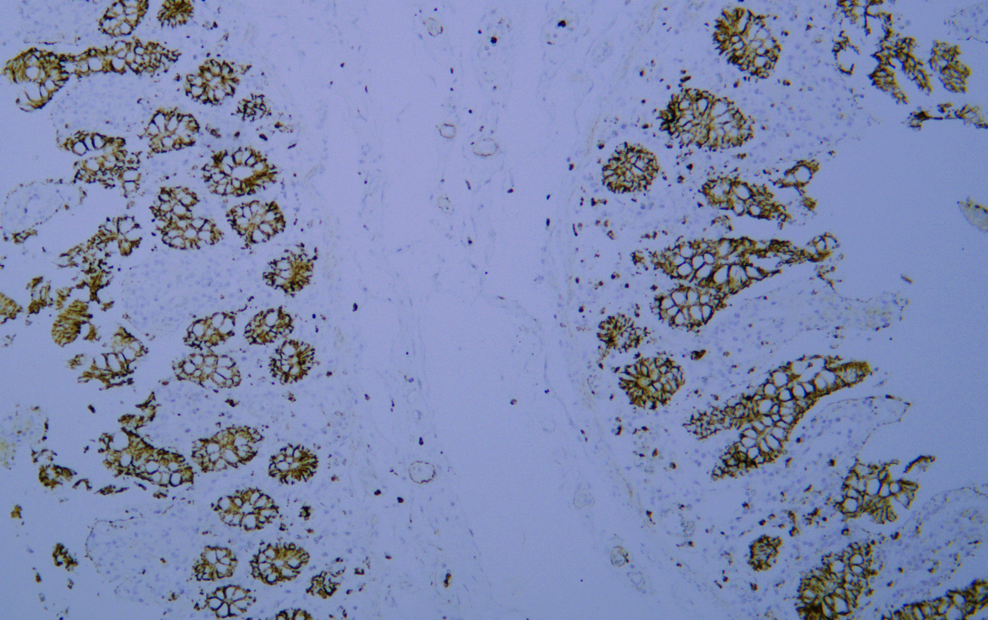
poly-ihc-rat-kidney.jpg)
- Immunohistochemical analysis of paraffin-embedded Rat-kidney tissue. 1,TGFβ RI (phospho Ser165) Polyclonal Antibody was diluted at 1:200(4°C,overnight). 2, Sodium citrate pH 6.0 was used for antibody retrieval(>98°C,20min). 3,Secondary antibody was diluted at 1:200(room tempeRature, 30min). Negative control was used by secondary antibody only.
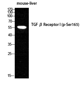
- Western Blot analysis of MOUSE-LIVER cells using Phospho-TGFβ RI (S165) Polyclonal Antibody diluted at 1:1000
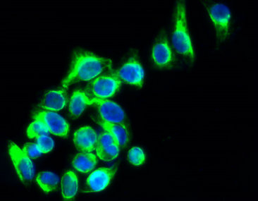
- Immunofluorescence analysis of HepG2 cells, Antibody diluted at 1:50. The picture on the right is blocked with the synthesized peptide.