Bax (phospho Thr167) Polyclonal Antibody
- Catalog No.:YP1169
- Applications:IF;ELISA
- Reactivity:Human;Mouse;Rat
- Target:
- Bax
- Fields:
- >>EGFR tyrosine kinase inhibitor resistance;>>Endocrine resistance;>>Platinum drug resistance;>>Sphingolipid signaling pathway;>>p53 signaling pathway;>>Protein processing in endoplasmic reticulum;>>Apoptosis;>>Longevity regulating pathway;>>Apoptosis - multiple species;>>Necroptosis;>>Neurotrophin signaling pathway;>>Non-alcoholic fatty liver disease;>>AGE-RAGE signaling pathway in diabetic complications;>>Parkinson disease;>>Amyotrophic lateral sclerosis;>>Huntington disease;>>Prion disease;>>Pathways of neurodegeneration - multiple diseases;>>Pathogenic Escherichia coli infection;>>Shigellosis;>>Salmonella infection;>>Tuberculosis;>>Hepatitis C;>>Hepatitis B;>>Measles;>>Human cytomegalovirus infection;>>Influenza A;>>Human papillomavirus infection;>>Human T-cell leukemia virus 1 infection;>>Kaposi sarcoma-associated herpesvirus infection;>>Herpes simplex virus 1 infection;>>Epstein-Barr virus infection;>>Human immunodeficiency virus 1 infection;>>Pathways in cancer;>>Transcriptional
- Gene Name:
- BAX
- Protein Name:
- Apoptosis regulator BAX
- Human Gene Id:
- 581
- Human Swiss Prot No:
- Q07812
- Mouse Gene Id:
- 12028
- Mouse Swiss Prot No:
- Q07813
- Rat Swiss Prot No:
- Q63690
- Immunogen:
- The antiserum was produced against synthesized peptide derived from human Bax around the phosphorylation site of Thr167. AA range:133-182
- Specificity:
- Phospho-Bax (T167) Polyclonal Antibody detects endogenous levels of Bax protein only when phosphorylated at T167.
- Formulation:
- Liquid in PBS containing 50% glycerol, 0.5% BSA and 0.02% sodium azide.
- Source:
- Polyclonal, Rabbit,IgG
- Dilution:
- IF 1:200 - 1:1000. ELISA: 1:40000. Not yet tested in other applications.
- Purification:
- The antibody was affinity-purified from rabbit antiserum by affinity-chromatography using epitope-specific immunogen.
- Concentration:
- 1 mg/ml
- Storage Stability:
- -15°C to -25°C/1 year(Do not lower than -25°C)
- Other Name:
- BAX;BCL2L4;Apoptosis regulator BAX;Bcl-2-like protein 4;Bcl2-L-4
- Molecular Weight(Da):
- 21kD
- Background:
- The protein encoded by BAX (BCL2 associated X, apoptosis regulator) belongs to the BCL2 protein family. BCL2 family members form hetero- or homodimers and act as anti- or pro-apoptotic regulators that are involved in a wide variety of cellular activities. This protein forms a heterodimer with BCL2, and functions as an apoptotic activator. This protein is reported to interact with, and increase the opening of, the mitochondrial voltage-dependent anion channel (VDAC), which leads to the loss in membrane potential and the release of cytochrome c. The expression of this gene is regulated by the tumor suppressor P53 and has been shown to be involved in P53-mediated apoptosis. Multiple alternatively spliced transcript variants, which encode different isoforms, have been reported for BAX.
- Function:
- disease:Defects in BAX are found in some cell lines from hematopoietic malignancies as T-cell acute lymphoblastic leukemia, Burkitt lymphoma, and plasmacytoma.,domain:Intact BH3 motif is required by BIK, BID, BAK, BAD and BAX for their pro-apoptotic activity and for their interaction with anti-apoptotic members of the Bcl-2 family.,function:Accelerates programmed cell death by binding to, and antagonizing the apoptosis repressor BCL2 or its adenovirus homolog E1B 19k protein. Induces the release of cytochrome c, activation of CASP3, and thereby apoptosis.,similarity:Belongs to the Bcl-2 family.,subcellular location:Colocalizes with 14-3-3 proteins in the cytoplasm. Under stress conditions, redistributes to the mitochondrion membrane through the release from JNK-phosphorylated 14-3-3 proteins.,subunit:Homodimer. Forms heterodimers with BCL2, E1B 19K protein, BCL2L1 isoform Bcl-X(L), MCL1
- Subcellular Location:
- [Isoform Alpha]: Mitochondrion outer membrane ; Single-pass membrane protein . Cytoplasm . Colocalizes with 14-3-3 proteins in the cytoplasm. Under stress conditions, undergoes a conformation change that causes release from JNK-phosphorylated 14-3-3 proteins and translocation to the mitochondrion membrane. Upon Sendai virus infection, recruited to the mitochondrion through interaction with IRF3 (PubMed:25609812). .; [Isoform Beta]: Cytoplasm.; [Isoform Gamma]: Cytoplasm.; [Isoform Delta]: Cytoplasm .
- Expression:
- Expressed in a wide variety of tissues. Isoform Psi is found in glial tumors. Isoform Alpha is expressed in spleen, breast, ovary, testis, colon and brain, and at low levels in skin and lung. Isoform Sigma is expressed in spleen, breast, ovary, testis, lung, colon, brain and at low levels in skin. Isoform Alpha and isoform Sigma are expressed in pro-myelocytic leukemia, histiocytic lymphoma, Burkitt's lymphoma, T-cell lymphoma, lymphoblastic leukemia, breast adenocarcinoma, ovary adenocarcinoma, prostate carcinoma, prostate adenocarcinoma, lung carcinoma, epidermoid carcinoma, small cell lung carcinoma and colon adenocarcinoma cell lines.
- June 19-2018
- WESTERN IMMUNOBLOTTING PROTOCOL
- June 19-2018
- IMMUNOHISTOCHEMISTRY-PARAFFIN PROTOCOL
- June 19-2018
- IMMUNOFLUORESCENCE PROTOCOL
- September 08-2020
- FLOW-CYTOMEYRT-PROTOCOL
- May 20-2022
- Cell-Based ELISA│解您多样本WB检测之困扰
- July 13-2018
- CELL-BASED-ELISA-PROTOCOL-FOR-ACETYL-PROTEIN
- July 13-2018
- CELL-BASED-ELISA-PROTOCOL-FOR-PHOSPHO-PROTEIN
- July 13-2018
- Antibody-FAQs
- Products Images

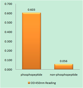
- Enzyme-Linked Immunosorbent Assay (Phospho-ELISA) for Immunogen Phosphopeptide (Phospho-left) and Non-Phosphopeptide (Phospho-right), using Bax (Phospho-Thr167) Antibody

- Immunofluorescence analysis of A549 cells, using Bax (Phospho-Thr167) Antibody. The picture on the right is blocked with the phospho peptide.


