Tak1 (phospho Thr184) Polyclonal Antibody
- Catalog No.:YP0378
- Applications:WB;IHC;IF;ELISA
- Reactivity:Human;Mouse;Rat
- Target:
- Tak1
- Fields:
- >>MAPK signaling pathway;>>NF-kappa B signaling pathway;>>Autophagy - animal;>>AMPK signaling pathway;>>Wnt signaling pathway;>>Osteoclast differentiation;>>Adherens junction;>>Neutrophil extracellular trap formation;>>Toll-like receptor signaling pathway;>>NOD-like receptor signaling pathway;>>RIG-I-like receptor signaling pathway;>>IL-17 signaling pathway;>>T cell receptor signaling pathway;>>TNF signaling pathway;>>Alcoholic liver disease;>>Pathogenic Escherichia coli infection;>>Shigellosis;>>Salmonella infection;>>Yersinia infection;>>Leishmaniasis;>>Toxoplasmosis;>>Hepatitis B;>>Measles;>>Herpes simplex virus 1 infection;>>Epstein-Barr virus infection;>>Human immunodeficiency virus 1 infection;>>Coronavirus disease - COVID-19;>>Lipid and atherosclerosis;>>Fluid shear stress and atherosclerosis
- Gene Name:
- MAP3K7
- Protein Name:
- Mitogen-activated protein kinase kinase kinase 7
- Human Gene Id:
- 6885
- Human Swiss Prot No:
- O43318
- Mouse Gene Id:
- 26409
- Mouse Swiss Prot No:
- Q62073
- Rat Gene Id:
- 1.00911e+008
- Rat Swiss Prot No:
- P0C8E4
- Immunogen:
- The antiserum was produced against synthesized peptide derived from human TAK1 around the phosphorylation site of Thr184. AA range:161-210
- Specificity:
- Phospho-Tak1 (T184) Polyclonal Antibody detects endogenous levels of Tak1 protein only when phosphorylated at T184.
- Formulation:
- Liquid in PBS containing 50% glycerol, 0.5% BSA and 0.02% sodium azide.
- Source:
- Polyclonal, Rabbit,IgG
- Dilution:
- WB 1:500 - 1:2000. IHC 1:100 - 1:300. ELISA: 1:10000.. IF 1:50-200
- Purification:
- The antibody was affinity-purified from rabbit antiserum by affinity-chromatography using epitope-specific immunogen.
- Concentration:
- 1 mg/ml
- Storage Stability:
- -15°C to -25°C/1 year(Do not lower than -25°C)
- Other Name:
- MAP3K7;TAK1;Mitogen-activated protein kinase kinase kinase 7;Transforming growth factor-beta-activated kinase 1;TGF-beta-activated kinase 1
- Observed Band(KD):
- 77kD
- Background:
- The protein encoded by this gene is a member of the serine/threonine protein kinase family. This kinase mediates the signaling transduction induced by TGF beta and morphogenetic protein (BMP), and controls a variety of cell functions including transcription regulation and apoptosis. In response to IL-1, this protein forms a kinase complex including TRAF6, MAP3K7P1/TAB1 and MAP3K7P2/TAB2; this complex is required for the activation of nuclear factor kappa B. This kinase can also activate MAPK8/JNK, MAP2K4/MKK4, and thus plays a role in the cell response to environmental stresses. Four alternatively spliced transcript variants encoding distinct isoforms have been reported. [provided by RefSeq, Jul 2008],
- Function:
- catalytic activity:ATP + a protein = ADP + a phosphoprotein.,cofactor:Magnesium.,function:Component of a protein kinase signal transduction cascade. Mediator of TGF-beta signal transduction. Stimulates NF-kappa-B activation and the p38 MAPK pathway.,PTM:Association with MAP3K7IP1 promotes autophosphorylation and subsequent activation. Dephosphorylation at Thr-187 by PP2A and PPP6C leads to inactivation.,similarity:Belongs to the protein kinase superfamily.,similarity:Belongs to the protein kinase superfamily. STE Ser/Thr protein kinase family. MAP kinase kinase kinase subfamily.,similarity:Contains 1 protein kinase domain.,subunit:Binds both upstream activators and downstream substrates in multimolecular complexes. Interacts with MAP3K7IP1 and MAP3K7IP2. Interacts with PPM1L. Interaction with PP2A and PPP6C leads to its' repressed activity.,
- Subcellular Location:
- Cytoplasm . Cell membrane ; Peripheral membrane protein ; Cytoplasmic side . Although the majority of MAP3K7/TAK1 is found in the cytosol, when complexed with TAB1/MAP3K7IP1 and TAB2/MAP3K7IP2, it is also localized at the cell membrane.
- Expression:
- Isoform 1A is the most abundant in ovary, skeletal muscle, spleen and blood mononuclear cells. Isoform 1B is highly expressed in brain, kidney and small intestine. Isoform 1C is the major form in prostate. Isoform 1D is the less abundant form.
- June 19-2018
- WESTERN IMMUNOBLOTTING PROTOCOL
- June 19-2018
- IMMUNOHISTOCHEMISTRY-PARAFFIN PROTOCOL
- June 19-2018
- IMMUNOFLUORESCENCE PROTOCOL
- September 08-2020
- FLOW-CYTOMEYRT-PROTOCOL
- May 20-2022
- Cell-Based ELISA│解您多样本WB检测之困扰
- July 13-2018
- CELL-BASED-ELISA-PROTOCOL-FOR-ACETYL-PROTEIN
- July 13-2018
- CELL-BASED-ELISA-PROTOCOL-FOR-PHOSPHO-PROTEIN
- July 13-2018
- Antibody-FAQs
- Products Images
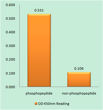
- Enzyme-Linked Immunosorbent Assay (Phospho-ELISA) for Immunogen Phosphopeptide (Phospho-left) and Non-Phosphopeptide (Phospho-right), using TAK1 (Phospho-Thr184) Antibody
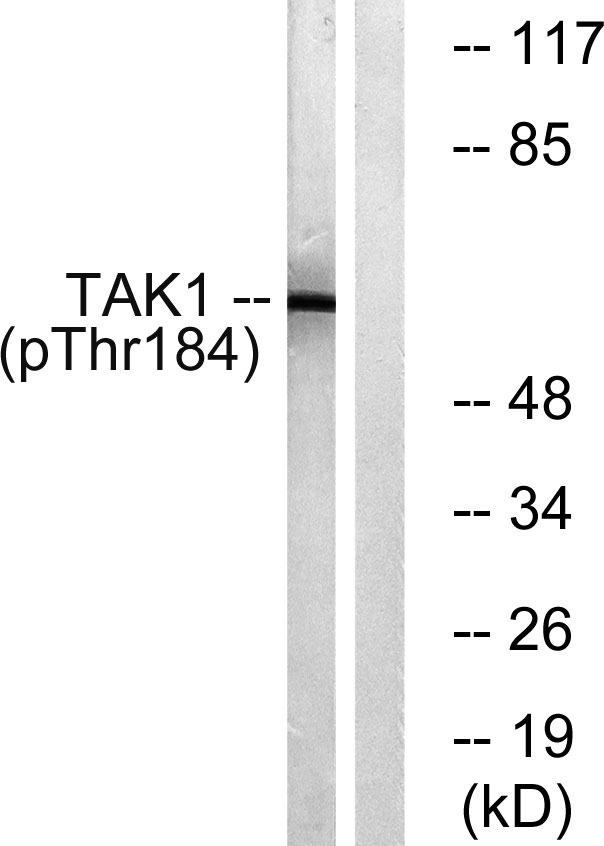
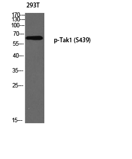
- Western blot analysis of lysates from HepG2 cells treated with TNF 20ng/ml 5', using TAK1 (Phospho-Thr184) Antibody. The lane on the right is blocked with the phospho peptide.
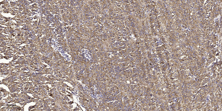
- Immunohistochemical analysis of paraffin-embedded human small intestinal carcinoma tissue. 1,primary Antibody was diluted at 1:200(4° overnight). 2, Sodium citrate pH 6.0 was used for antigen retrieval(>98°C,20min). 3,Secondary antibody was diluted at 1:200

.jpg)