CD4 (PT0412R) PT® Rabbit mAb
- Catalog No.:YM8254
- Applications:WB;IHC;IF;IP;ELISA
- Reactivity:Human;
- Target:
- CD4
- Gene Name:
- CD4
- Protein Name:
- T-cell surface glycoprotein CD4 (T-cell surface antigen T4/Leu-3) (CD antigen CD4)
- Human Gene Id:
- 920
- Human Swiss Prot No:
- P01730
- Mouse Gene Id:
- 12504
- Mouse Swiss Prot No:
- P06332
- Rat Gene Id:
- 24932
- Rat Swiss Prot No:
- P05540
- Specificity:
- endogenous
- Formulation:
- PBS, 50% glycerol, 0.05% Proclin 300, 0.05%BSA
- Source:
- Monoclonal, rabbit, IgG, Kappa
- Dilution:
- IHC 1:1000-1:5000;WB 1:1000-1:5000;IF 1:200-1:1000;ELISA 1:5000-1:20000;IP 1:50-1:200;
- Purification:
- Protein A
- Storage Stability:
- -15°C to -25°C/1 year(Do not lower than -25°C)
- Other Name:
- T-cell surface glycoprotein CD4 (T-cell surface antigen T4/Leu-3) (CD antigen CD4)
- Molecular Weight(Da):
- 51kD
- Observed Band(KD):
- 55kD
- Background:
- CD4 molecule(CD4) Homo sapiens This gene encodes a membrane glycoprotein of T lymphocytes that interacts with major histocompatibility complex class II antigenes and is also a receptor for the human immunodeficiency virus. This gene is expressed not only in T lymphocytes, but also in B cells, macrophages, and granulocytes. It is also expressed in specific regions of the brain. The protein functions to initiate or augment the early phase of T-cell activation, and may function as an important mediator of indirect neuronal damage in infectious and immune-mediated diseases of the central nervous system. Multiple alternatively spliced transcript variants encoding different isoforms have been identified in this gene. [provided by RefSeq, Aug 2010],
- Function:
- Integral membrane glycoprotein that plays an essential role in the immune response and serves multiple functions in responses against both external and internal offenses. In T-cells, functions primarily as a coreceptor for MHC class II molecule:peptide complex. The antigens presented by class II peptides are derived from extracellular proteins while class I peptides are derived from cytosolic proteins. Interacts simultaneously with the T-cell receptor (TCR) and the MHC class II presented by antigen presenting cells (APCs). In turn, recruits the Src kinase LCK to the vicinity of the TCR-CD3 complex. LCK then initiates different intracellular signaling pathways by phosphorylating various substrates ultimately leading to lymphokine production, motility, adhesion and activation of T-helper cells. In other cells such as macrophages or NK cells, plays a role in differentiation/activation, cyt
- Subcellular Location:
- Membrane
- Expression:
- Highly expressed in T-helper cells. The presence of CD4 is a hallmark of T-helper cells which are specialized in the activation and growth of cytotoxic T-cells, regulation of B cells, or activation of phagocytes. CD4 is also present in other immune cells such as macrophages, dendritic cells or NK cells.
- June 19-2018
- WESTERN IMMUNOBLOTTING PROTOCOL
- June 19-2018
- IMMUNOHISTOCHEMISTRY-PARAFFIN PROTOCOL
- June 19-2018
- IMMUNOFLUORESCENCE PROTOCOL
- September 08-2020
- FLOW-CYTOMEYRT-PROTOCOL
- May 20-2022
- Cell-Based ELISA│解您多样本WB检测之困扰
- July 13-2018
- CELL-BASED-ELISA-PROTOCOL-FOR-ACETYL-PROTEIN
- July 13-2018
- CELL-BASED-ELISA-PROTOCOL-FOR-PHOSPHO-PROTEIN
- July 13-2018
- Antibody-FAQs
- Products Images
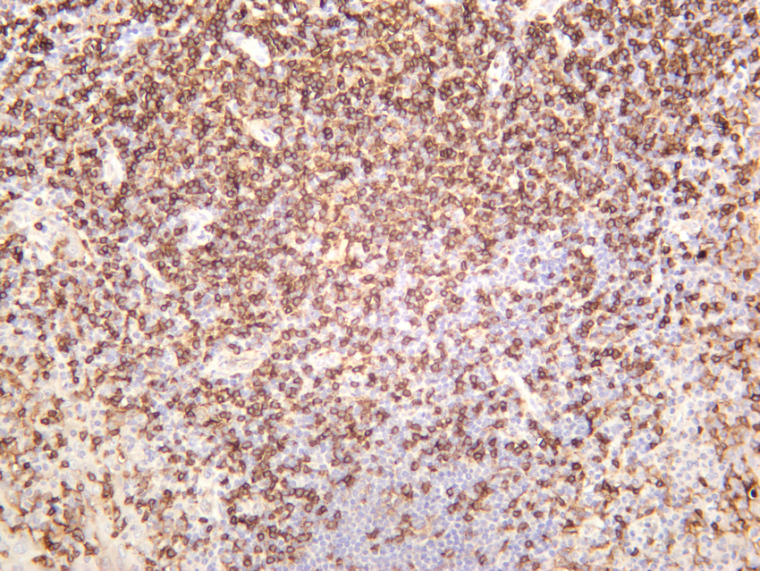
- Human tonsil was stained with anti-CD4 (PT0412R) rabbit antibody
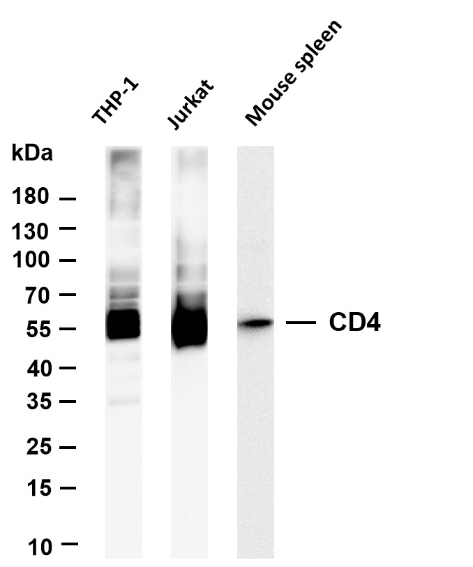
- Various whole cell lysates were separated by 4-20% SDS-PAGE, and the membrane was blotted with anti-CD4 (PT0412R) antibody. The HRP-conjugated Goat anti-Rabbit IgG(H + L) antibody was used to detect the antibody. Lane 1: THP-1 Lane 2: Jurkat Lane 2: Mouse spleen Predicted band size: 51kDa Observed band size: 51kDa
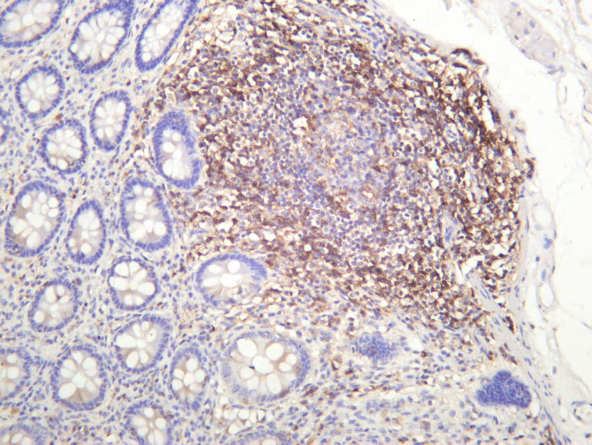
- Human colon was stained with anti-CD4 (PT0412R) rabbit antibody
