HDAC1 (PT0390R) PT® Rabbit mAb
- Catalog No.:YM8239
- Applications:WB;IHC;IF;IP;ELISA
- Reactivity:Human; Mouse; Rat;
- Target:
- HDAC1
- Fields:
- >>Cell cycle;>>Longevity regulating pathway - multiple species;>>Notch signaling pathway;>>Neutrophil extracellular trap formation;>>Thyroid hormone signaling pathway;>>Huntington disease;>>Amphetamine addiction;>>Alcoholism;>>Human papillomavirus infection;>>Epstein-Barr virus infection;>>Pathways in cancer;>>Transcriptional misregulation in cancer;>>Viral carcinogenesis;>>MicroRNAs in cancer;>>Chronic myeloid leukemia
- Gene Name:
- HDAC1
- Protein Name:
- Histone deacetylase 1
- Human Gene Id:
- 3065
- Human Swiss Prot No:
- Q13547
- Mouse Gene Id:
- 433759
- Mouse Swiss Prot No:
- O09106
- Rat Gene Id:
- 297893
- Rat Swiss Prot No:
- Q4QQW4
- Specificity:
- endogenous
- Formulation:
- PBS, 50% glycerol, 0.05% Proclin 300, 0.05%BSA
- Source:
- Monoclonal, rabbit, IgG, Kappa
- Dilution:
- IHC 1:4000-1:10000,WB 1:1000-1:5000,IF 1:200-1:1000,ELISA 1:5000-1:20000,IP 1:50-1:200,
- Purification:
- Protein A
- Storage Stability:
- -15°C to -25°C/1 year(Do not lower than -25°C)
- Other Name:
- HDAC1;RPD3L1;Histone deacetylase 1;HD1
- Molecular Weight(Da):
- 55kD
- Observed Band(KD):
- 62kD
- Background:
- Histone acetylation and deacetylation, catalyzed by multisubunit complexes, play a key role in the regulation of eukaryotic gene expression. The protein encoded by this gene belongs to the histone deacetylase/acuc/apha family and is a component of the histone deacetylase complex. It also interacts with retinoblastoma tumor-suppressor protein and this complex is a key element in the control of cell proliferation and differentiation. Together with metastasis-associated protein-2, it deacetylates p53 and modulates its effect on cell growth and apoptosis. [provided by RefSeq, Jul 2008],
- Function:
- catalytic activity:Hydrolysis of an N(6)-acetyl-lysine residue of a histone to yield a deacetylated histone.,function:Responsible for the deacetylation of lysine residues on the N-terminal part of the core histones (H2A, H2B, H3 and H4). Histone deacetylation gives a tag for epigenetic repression and plays an important role in transcriptional regulation, cell cycle progression and developmental events. Histone deacetylases act via the formation of large multiprotein complexes.,PTM:Phosphorylation on Ser-421 and Ser-423 promotes enzymatic activity and interactions with NuRD and SIN3 complexes.,PTM:Sumoylated on Lys-444 and Lys-476; which promotes enzymatic activity. Desumoylated by SENP1.,similarity:Belongs to the histone deacetylase family. Type 1 subfamily.,subunit:Part of the core histone deacetylase (HDAC) complex composed of HDAC1, HDAC2, RBBP4 and RBBP7. The core complex associates
- Subcellular Location:
- Nucleus
- Expression:
- Ubiquitous, with higher levels in heart, pancreas and testis, and lower levels in kidney and brain.
MiR-548an, Transcriptionally Downregulated by HIF1α/HDAC1, Suppresses Tumorigenesis of Pancreatic Cancer by Targeting Vimentin Expression. MOLECULAR CANCER THERAPEUTICS Mol Cancer Ther. 2016 Sep;15(9):2209-2219 WB Human 1:1000 BxPC-3 cell
Panobinostat reverses HepaCAM gene expression and suppresses proliferation by increasing histone acetylation in prostate cancer. GENE Gene. 2022 Jan;808:145977 WB Human LNCaP cell,DU145 cell
MiR-548an, transcriptionally downregulated by HIF-1α/HDAC1, suppresses tumorigenesis of pancreatic cancer by targeting vimentin expression." Molecular cancer therapeutics (2016): molcanther-0877.
A di-acetyl-decorated chromatin signature couples liquid condensation to suppress DNA end synapsis MOLECULAR CELL Kaiwen Bao WB,CoIP Human HeLa cell
- June 19-2018
- WESTERN IMMUNOBLOTTING PROTOCOL
- June 19-2018
- IMMUNOHISTOCHEMISTRY-PARAFFIN PROTOCOL
- June 19-2018
- IMMUNOFLUORESCENCE PROTOCOL
- September 08-2020
- FLOW-CYTOMEYRT-PROTOCOL
- May 20-2022
- Cell-Based ELISA│解您多样本WB检测之困扰
- July 13-2018
- CELL-BASED-ELISA-PROTOCOL-FOR-ACETYL-PROTEIN
- July 13-2018
- CELL-BASED-ELISA-PROTOCOL-FOR-PHOSPHO-PROTEIN
- July 13-2018
- Antibody-FAQs
- Products Images

- Various whole cell lysates were separated by 4-20% SDS-PAGE, and the membrane was blotted with anti-HDAC1 (PT0390R) antibody. The HRP-conjugated Goat anti-Rabbit IgG(H + L) antibody was used to detect the antibody. Lane 1: K562 Lane 2: Hela Lane 3: NIH-3T3 Lane 4: C6 Predicted band size: 55kDa Observed band size: 62kDa

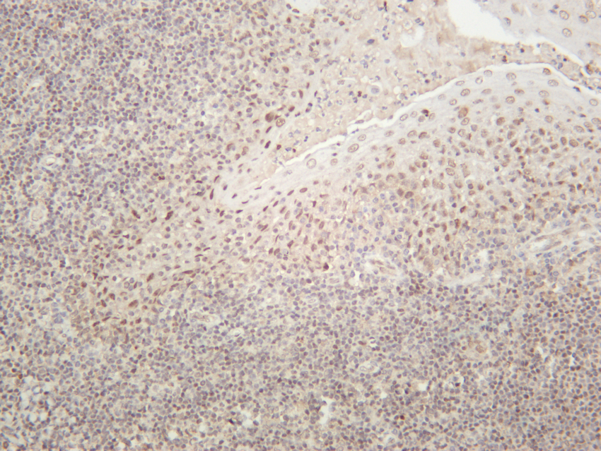
- Human tonsil was stained with anti-HDAC1 (PT0390R) rabbit antibody

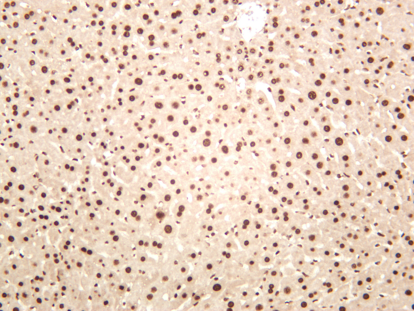
- Mouse liver was stained with anti-HDAC1 (PT0390R) rabbit antibody

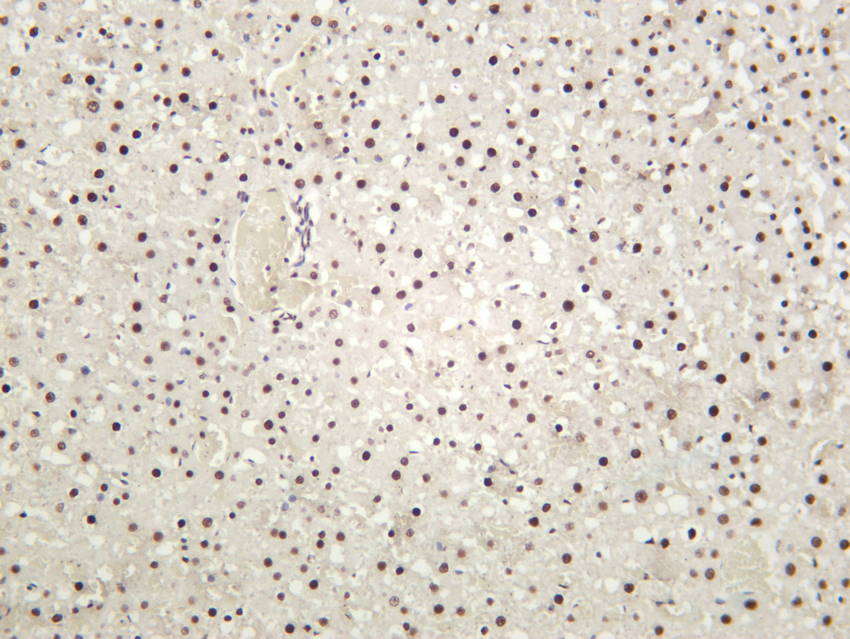
- Rat liver was stained with anti-HDAC1 (PT0390R) rabbit antibody


