CD38 (PT0361R) PT® Rabbit mAb
- Catalog No.:YM8214
- Applications:WB;IHC;IF;IP;ELISA
- Reactivity:Human;
- Target:
- CD38
- Fields:
- >>Nicotinate and nicotinamide metabolism;>>Metabolic pathways;>>Calcium signaling pathway;>>Hematopoietic cell lineage;>>Oxytocin signaling pathway;>>Salivary secretion;>>Pancreatic secretion
- Gene Name:
- CD38
- Protein Name:
- ADP-ribosyl cyclase 1
- Human Gene Id:
- 952
- Human Swiss Prot No:
- P28907
- Mouse Swiss Prot No:
- P56528
- Specificity:
- endogenous
- Formulation:
- PBS, 50% glycerol, 0.05% Proclin 300, 0.05%BSA
- Source:
- Monoclonal, rabbit, IgG, Kappa
- Dilution:
- IHC 1:10000-1:30000;WB 1:1000-1:5000;IF 1:200-1:1000;ELISA 1:5000-1:20000;IP 1:50-1:200;
- Purification:
- Protein A
- Storage Stability:
- -15°C to -25°C/1 year(Do not lower than -25°C)
- Other Name:
- CD38;ADP-ribosyl cyclase 1;Cyclic ADP-ribose hydrolase 1;cADPr hydrolase 1;T10;CD antigen CD38
- Molecular Weight(Da):
- 34kD
- Observed Band(KD):
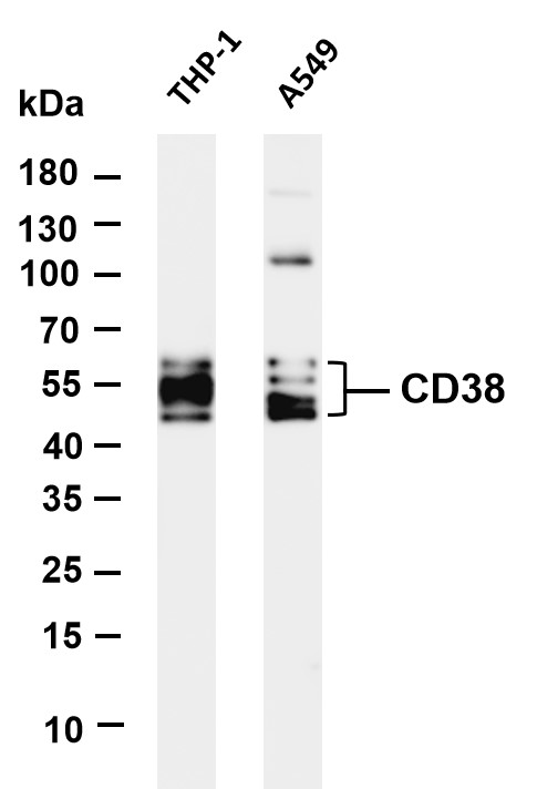
- 45-60kD
- Background:
- The protein encoded by this gene is a non-lineage-restricted, type II transmembrane glycoprotein that synthesizes and hydrolyzes cyclic adenosine 5'-diphosphate-ribose, an intracellular calcium ion mobilizing messenger. The release of soluble protein and the ability of membrane-bound protein to become internalized indicate both extracellular and intracellular functions for the protein. This protein has an N-terminal cytoplasmic tail, a single membrane-spanning domain, and a C-terminal extracellular region with four N-glycosylation sites. Crystal structure analysis demonstrates that the functional molecule is a dimer, with the central portion containing the catalytic site. It is used as a prognostic marker for patients with chronic lymphocytic leukemia. Alternative splicing results in multiple transcript variants. [provided by RefSeq, Sep 2015],
- Function:
- catalytic activity:NAD(+) + H(2)O = ADP-ribose + nicotinamide.,developmental stage:Preferentially expressed at both early and late stages of the B and T-cell maturation. It is also detected on erythroid and myeloid progenitors in bone marrow, where the level of surface expression was shown to decrease during differentiation of blast-forming unit E to colony-forming unit E.,enzyme regulation:ATP inhibits the hydrolyzing activity.,function:Synthesizes cyclic ADP-ribose, a second messenger for glucose-induced insulin secretion. Also has cADPr hydrolase activity. Also moonlights as a receptor in cells of the immune system.,online information:CD38 entry,similarity:Belongs to the ADP-ribosyl cyclase family.,tissue specificity:Expressed at high levels in pancreas, liver, kidney, brain, testis, ovary, placenta, malignant lymphoma and neuroblastoma.,
- Subcellular Location:
- Membrane
- Expression:
- Expressed at high levels in pancreas, liver, kidney, brain, testis, ovary, placenta, malignant lymphoma and neuroblastoma.
- June 19-2018
- WESTERN IMMUNOBLOTTING PROTOCOL
- June 19-2018
- IMMUNOHISTOCHEMISTRY-PARAFFIN PROTOCOL
- June 19-2018
- IMMUNOFLUORESCENCE PROTOCOL
- September 08-2020
- FLOW-CYTOMEYRT-PROTOCOL
- May 20-2022
- Cell-Based ELISA│解您多样本WB检测之困扰
- July 13-2018
- CELL-BASED-ELISA-PROTOCOL-FOR-ACETYL-PROTEIN
- July 13-2018
- CELL-BASED-ELISA-PROTOCOL-FOR-PHOSPHO-PROTEIN
- July 13-2018
- Antibody-FAQs
- Products Images

- Various whole cell lysates were separated by 4-20% SDS-PAGE, and the membrane was blotted with anti-CD38 (PT0361R) antibody. The HRP-conjugated Goat anti-Rabbit IgG(H + L) antibody was used to detect the antibody. Lane 1: THP-1 Lane 2: A549 Predicted band size: 34kDa Observed band size: 45-65kDa

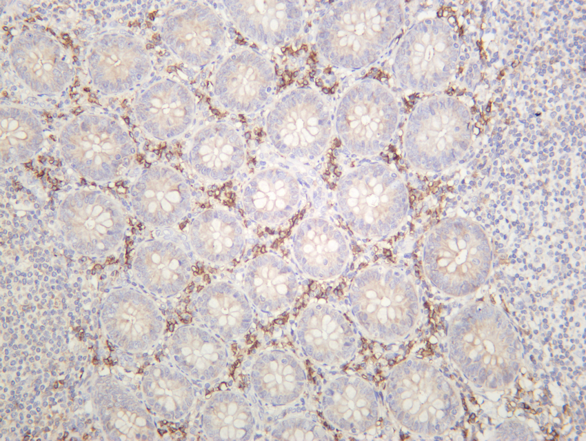
- Human appendix was stained with anti-CD38 (PT0361R) rabbit antibody

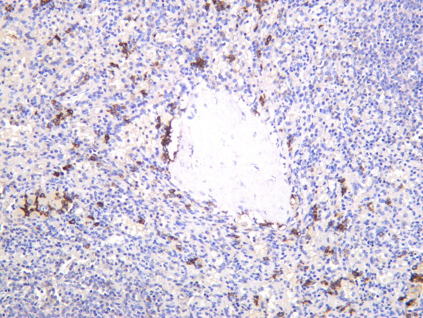
- Human spleen was stained with anti-CD38 (PT0361R) rabbit antibody


