VWF (PT0316R) PT® Rabbit mAb
- Catalog No.:YM8187
- Applications:WB;IHC;IF;IP;ELISA
- Reactivity:Human; Mouse; Rat;
- Target:
- VWF
- Fields:
- >>PI3K-Akt signaling pathway;>>Focal adhesion;>>ECM-receptor interaction;>>Complement and coagulation cascades;>>Platelet activation;>>Neutrophil extracellular trap formation;>>Human papillomavirus infection;>>Coronavirus disease - COVID-19
- Gene Name:
- VWF F8VWF
- Protein Name:
- von Willebrand factor (vWF) [Cleaved into: von Willebrand antigen 2 (von Willebrand antigen II)]
- Human Gene Id:
- 7450
- Human Swiss Prot No:
- P04275
- Mouse Swiss Prot No:
- Q8CIZ8
- Rat Swiss Prot No:
- Q62935
- Specificity:
- endogenous
- Formulation:
- PBS, 50% glycerol, 0.05% Proclin 300, 0.05%BSA
- Source:
- Monoclonal, rabbit, IgG, Kappa
- Dilution:
- IHC 1:1000-1:4000;WB 1:2000-1:10000;IF 1:200-1:1000;ELISA 1:5000-1:20000;IP 1:50-1:200;
- Purification:
- Protein A
- Storage Stability:
- -15°C to -25°C/1 year(Do not lower than -25°C)
- Molecular Weight(Da):
- 309kD
- Observed Band(KD):
- 280kD
- Background:
- This gene encodes a glycoprotein involved in hemostasis. The encoded preproprotein is proteolytically processed following assembly into large multimeric complexes. These complexes function in the adhesion of platelets to sites of vascular injury and the transport of various proteins in the blood. Mutations in this gene result in von Willebrand disease, an inherited bleeding disorder. An unprocessed pseudogene has been found on chromosome 22. [provided by RefSeq, Oct 2015],
- Function:
- disease:Defects in VWF are associated with various forms of von Willebrand disease (VWD) [MIM:193400, 277480]. VWD is characterized by frequent bleeding (gingival, minor skin quantitative lacerations, menorrhagia, etc.). Type I VWD is associated with a deficiency of VWF; type II by normal to decreased plasma level of VWF; type III by a virtual absence of VWF. There are subtypes (A to H) of type II VWD; for example: type IIA is characterized by the absence of VWF high molecular weight multimers in plasma.,domain:The von Willebrand antigen 2 is required for multimerization of vWF and for its targeting to storage granules.,function:Important in the maintenance of hemostasis, it promotes adhesion of platelets to the sites of vascular injury by forming a molecular bridge between sub-endothelial collagen matrix and platelet-surface receptor complex GPIb-IX-V. Also acts as a chaperone for coagu
- Subcellular Location:
- Secreted
- Expression:
- Plasma.
A novel leptin receptor binding peptide tethered-collagen scaffold promotes lung injury repair
Liu, Ting, Xuelian Du, and Xiugui Sheng. "Gene expression changes after ionizing radiation in endothelial cells derived from human endometrial cancer-preliminary outcomes." Archives of gynecology and obstetrics 289.6 (2014): 1315-1323.
Hui, Zhi, and Zhe Li. "Continuous veno-venous hemofiltration for septic shock." INTERNATIONAL JOURNAL OF CLINICAL AND EXPERIMENTAL MEDICINE 10.10 (2017): 14983-14988.
Lu, Xiangwei, et al. "Gene alterations after ionizing radiation in human cervical carcinoma-associated endothelial cells." Int J Clin Exp Pathol 9.2 (2016): 1093-1104.
司晓丽, et al. "在不同转移潜能人肺癌细胞移植瘤中血管性血友病因子的表达." 基础医学与临床 40.3 (2020): 346.
- June 19-2018
- WESTERN IMMUNOBLOTTING PROTOCOL
- June 19-2018
- IMMUNOHISTOCHEMISTRY-PARAFFIN PROTOCOL
- June 19-2018
- IMMUNOFLUORESCENCE PROTOCOL
- September 08-2020
- FLOW-CYTOMEYRT-PROTOCOL
- May 20-2022
- Cell-Based ELISA│解您多样本WB检测之困扰
- July 13-2018
- CELL-BASED-ELISA-PROTOCOL-FOR-ACETYL-PROTEIN
- July 13-2018
- CELL-BASED-ELISA-PROTOCOL-FOR-PHOSPHO-PROTEIN
- July 13-2018
- Antibody-FAQs
- Products Images

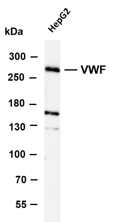
- Various whole cell lysates were separated by 4-8% SDS-PAGE, and the membrane was blotted with anti- VWF (PT0316R) antibody. The HRP-conjugated Goat anti-Rabbit IgG(H + L) antibody was used to detect the antibody. Lane 1: HepG2 Predicted band size: 309kDa Observed band size: 280kDa

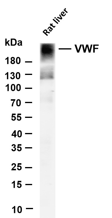
- Various whole cell lysates were separated by 4-20% SDS-PAGE, and the membrane was blotted with anti-VWF (PT0316R) antibody. The HRP-conjugated Goat anti-Rabbit IgG(H + L) antibody was used to detect the antibody. Lane 1: Rat liver Predicted band size: 309kDa Observed band size: 280kDa

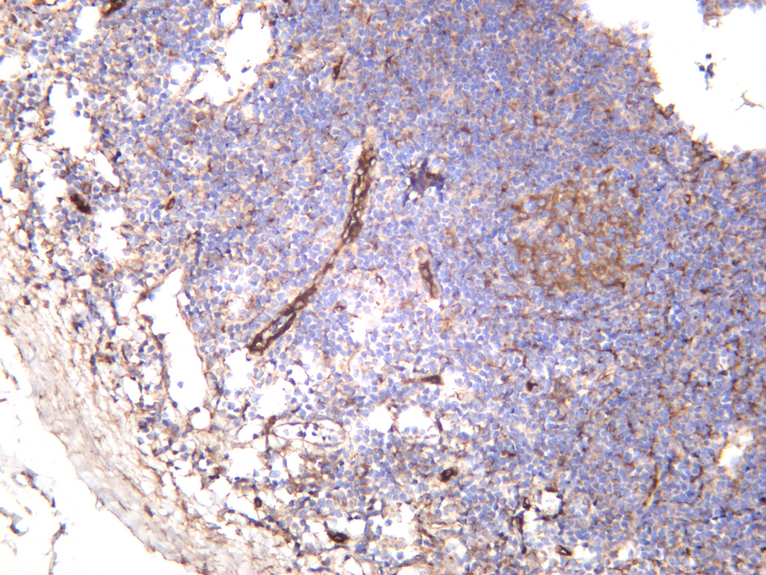
- Human tonsil was stained with anti-VWF (PT0316R) rabbit antibody

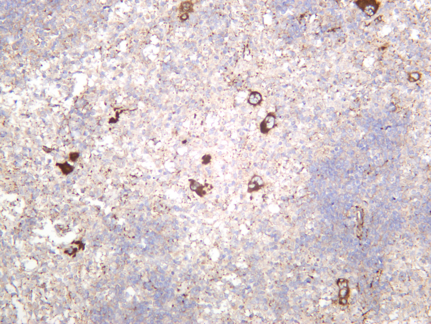
- Rat spleen was stained with anti-VWF (PT0316R) rabbit antibody


