CD86 (PT0048R) PT® Rabbit mAb
- Catalog No.:YM8023
- Applications:WB;IHC;IF;IP;ELISA
- Reactivity:Mouse;
- Target:
- CD86
- Fields:
- >>Cell adhesion molecules;>>Toll-like receptor signaling pathway;>>Intestinal immune network for IgA production;>>Type I diabetes mellitus;>>Kaposi sarcoma-associated herpesvirus infection;>>Transcriptional misregulation in cancer;>>Autoimmune thyroid disease;>>Systemic lupus erythematosus;>>Rheumatoid arthritis;>>Allograft rejection;>>Graft-versus-host disease;>>Viral myocarditis
- Gene Name:
- CD86 CD28LG2
- Protein Name:
- CD86
- Human Gene Id:
- 942
- Human Swiss Prot No:
- P42081
- Mouse Gene Id:
- 12524
- Mouse Swiss Prot No:
- P42082
- Specificity:
- endogenous
- Formulation:
- PBS, 50% glycerol, 0.05% Proclin 300, 0.05%BSA
- Source:
- Monoclonal, rabbit, IgG, Kappa
- Dilution:
- IHC 1:200-1000,WB 1:500-5000,IF 1:200-1000,ELISA 1:5000-20000,IP 1:50-200
- Purification:
- Protein A
- Storage Stability:
- -15°C to -25°C/1 year(Do not lower than -25°C)
- Other Name:
- T-lymphocyte activation antigen CD86 (Activation B7-2 antigen;B70;BU63;CTLA-4 counter-receptor B7.2;FUN-1;CD antigen CD86)
- Molecular Weight(Da):
- 60-80kD
- Background:
- This gene encodes a type I membrane protein that is a member of the immunoglobulin superfamily. This protein is expressed by antigen-presenting cells, and it is the ligand for two proteins at the cell surface of T cells, CD28 antigen and cytotoxic T-lymphocyte-associated protein 4. Binding of this protein with CD28 antigen is a costimulatory signal for activation of the T-cell. Binding of this protein with cytotoxic T-lymphocyte-associated protein 4 negatively regulates T-cell activation and diminishes the immune response. Alternative splicing results in several transcript variants encoding different isoforms.[provided by RefSeq, May 2011],
- Function:
- function:Receptor involved in the costimulatory signal essential for T-lymphocyte proliferation and interleukin-2 production, by binding CD28 or CTLA-4. May play a critical role in the early events of T-cell activation and costimulation of naive T-cells, such as deciding between immunity and anergy that is made by T-cells within 24 hours after activation. Isoform 2 interferes with the formation of CD86 clusters, and thus acts as a negative regulator of T-cell activation.,online information:CD86 entry,PTM:Polyubiquitinated; which is promoted by MARCH8 and results in endocytosis and lysosomal degradation.,similarity:Contains 1 Ig-like C2-type (immunoglobulin-like) domain.,similarity:Contains 1 Ig-like V-type (immunoglobulin-like) domain.,subunit:Interacts with MARCH8. Interacts with human herpesvirus 8 MIR2 protein (Probable). Interacts with adenovirus subgroup B fiber proteins and acts as
- Subcellular Location:
- Membranous
- June 19-2018
- WESTERN IMMUNOBLOTTING PROTOCOL
- June 19-2018
- IMMUNOHISTOCHEMISTRY-PARAFFIN PROTOCOL
- June 19-2018
- IMMUNOFLUORESCENCE PROTOCOL
- September 08-2020
- FLOW-CYTOMEYRT-PROTOCOL
- May 20-2022
- Cell-Based ELISA│解您多样本WB检测之困扰
- July 13-2018
- CELL-BASED-ELISA-PROTOCOL-FOR-ACETYL-PROTEIN
- July 13-2018
- CELL-BASED-ELISA-PROTOCOL-FOR-PHOSPHO-PROTEIN
- July 13-2018
- Antibody-FAQs
- Products Images

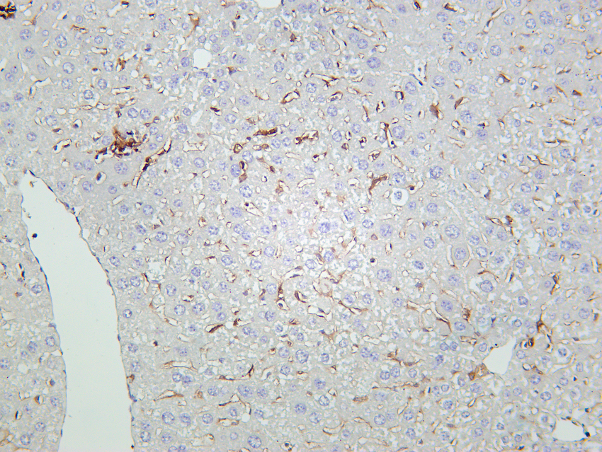
- Mouse liver tissue was stained with Anti-CD86 (PT0048R) rabbit Antibody

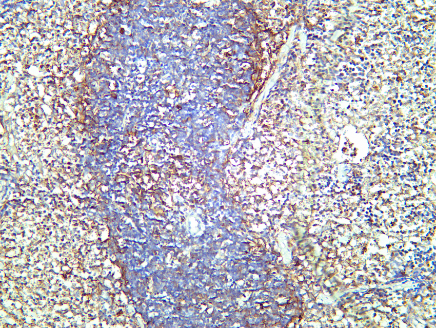
- Mouse spleen tissue was stained with Anti-CD86 (PT0048R) rabbit Antibody

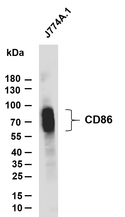
- Various whole cell lysates were separated by 4-20% SDS-PAGE, and the membrane was blotted with anti-CD86 (PT0048R) antibody. The HRP-conjugated Goat anti-Rabbit IgG(H + L) antibody was used to detect the antibody. Lane 1: J774A.1 Predicted band size: 35kDa Observed band size: 60-85kDa

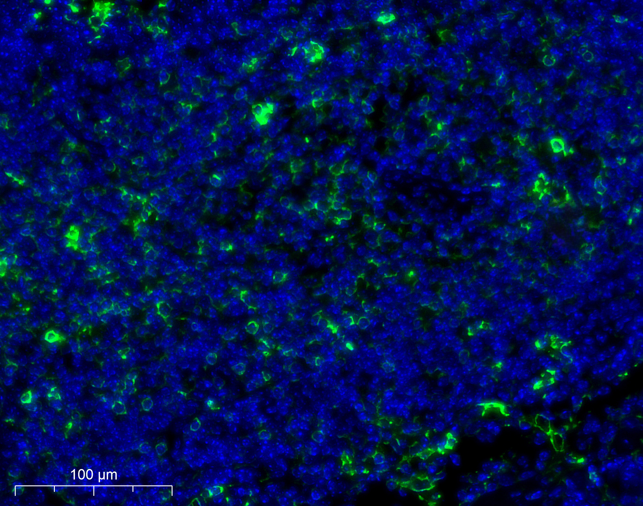
- Immunofluorescence analysis of paraffin-embedded Mouse spleen. Primary Antibody was diluted at 1:200(4° overnight). an Multi colour-Fluorescence kit (RS0035, Immunoway). EDTA based antigen retrieval was used before Green tyramide signal amplification. DAPI (dark blue) was used as a nuclear counter stain. Microscopy and pseudocoloring of individual dyes was performed using a Slideviewer Imaging System (3D histech).


