Cytokeratin 4 (ABT-CK4) mouse mAb
- Catalog No.:YM6038
- Applications:IHC;WB;ELISA
- Reactivity:Human
- Target:
- Cytokeratin 4
- Gene Name:
- KRT4 CYK4
- Protein Name:
- Keratin, type II cytoskeletal 4 (Cytokeratin-4) (CK-4) (Keratin-4) (K4) (Type-II keratin Kb4)
- Human Swiss Prot No:
- P19013
- Immunogen:
- Synthesized peptide derived from human Cytokeratin 4 AA range: 400-534
- Specificity:
- This antibody detects endogenous levels of human Cytokeratin 4. Heat-induced epitope retrieval (HIER) Citrate buffer of pH6.0 was highly recommended as antigen repair method in paraffin section
- Formulation:
- Liquid in PBS containing 50% glycerol, 0.5% BSA and 0.02% sodium azide.
- Source:
- Mouse, Monoclonal/IgG2a, Kappa
- Dilution:
- WB 1:500-2000 IHC 1:200-400, ELISA 1:5000-20000
- Purification:
- The antibody was affinity-purified from mouse ascites by affinity-chromatography using specific immunogen.
- Storage Stability:
- -15°C to -25°C/1 year(Do not lower than -25°C)
- Molecular Weight(Da):
- 56kD
- Background:
- keratin 4(KRT4) Homo sapiens The protein encoded by this gene is a member of the keratin gene family. The type II cytokeratins consist of basic or neutral proteins which are arranged in pairs of heterotypic keratin chains coexpressed during differentiation of simple and stratified epithelial tissues. This type II cytokeratin is specifically expressed in differentiated layers of the mucosal and esophageal epithelia with family member KRT13. Mutations in these genes have been associated with White Sponge Nevus, characterized by oral, esophageal, and anal leukoplakia. The type II cytokeratins are clustered in a region of chromosome 12q12-q13. [provided by RefSeq, Jul 2008],
- Function:
- disease:Defects in KRT4 are a cause of white sponge nevus of cannon (WSN) [MIM:193900]. WSN is a rare autosomal dominant disorder which predominantly affects non-cornified stratified squamous epithelia. Clinically, it is characterized by the presence of soft, white, and spongy plaques in the oral mucosa. The characteristic histopathologic features are epithelial thickening, parakeratosis, and vacuolization of the suprabasal layer of oral epithelial keratinocytes. Less frequently the mucous membranes of the nose, esophagus, genitalia and rectum are involved.,miscellaneous:There are two types of cytoskeletal and microfibrillar keratin: I (acidic; 40-55 kDa) and II (neutral to basic; 56-70 kDa).,polymorphism:Three alleles of K4 are known: K4A2 (shown here), K4A1 and K4B.,similarity:Belongs to the intermediate filament family.,subunit:Heterotetramer of two type I and two type II keratins. Ke
- Subcellular Location:
- Cytoplasmic, Membranous
- Expression:
- Detected in the suprabasal layer of the stratified epithelium of the esophagus, exocervix, vagina, mouth and lingual mucosa, and in cells and cell clusters in the mucosa and serous gland ducts of the esophageal submucosa (at protein level). Expressed widely in the exocervix and esophageal epithelium, with lowest levels detected in the basal cell layer.
- June 19-2018
- WESTERN IMMUNOBLOTTING PROTOCOL
- June 19-2018
- IMMUNOHISTOCHEMISTRY-PARAFFIN PROTOCOL
- June 19-2018
- IMMUNOFLUORESCENCE PROTOCOL
- September 08-2020
- FLOW-CYTOMEYRT-PROTOCOL
- May 20-2022
- Cell-Based ELISA│解您多样本WB检测之困扰
- July 13-2018
- CELL-BASED-ELISA-PROTOCOL-FOR-ACETYL-PROTEIN
- July 13-2018
- CELL-BASED-ELISA-PROTOCOL-FOR-PHOSPHO-PROTEIN
- July 13-2018
- Antibody-FAQs
- Products Images
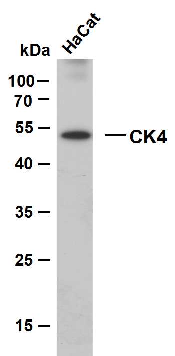
- HaCat whole cell lysates were separated by 10% SDS-PAGE, and the membrane was blotted with anti-CK4(ABT-CK4) antibody. The HRP-conjugated Goat anti-Mouse IgG(H + L) antibody was used to detect the antibody. Lane 1: HaCat Predicted band size: 57kDa Observed band size: 54kDa
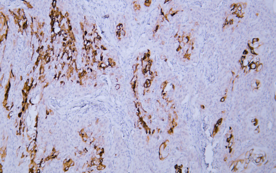
- Human cervical squamous cell carcinoma tissue was stained with Anti-Cytokeratin 4 (ABT-CK4) Antibody
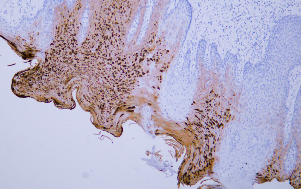
- Human oral squamous cell carcinoma tissue was stained with Anti-Cytokeratin 4 (ABT-CK4) Antibody


