MyoD1 (ABT-MYOD1) mouse mAb
- Catalog No.:YM4945
- Applications:IHC;IF;ELISA
- Reactivity:Human;Mouse;Rat;
- Target:
- MyoD
- Fields:
- >>Spinocerebellar ataxia
- Gene Name:
- MYOD1 BHLHC1 MYF3 MYOD
- Protein Name:
- Myoblast determination protein 1 (Class C basic helix-loop-helix protein 1) (bHLHc1) (Myogenic factor 3) (Myf-3)
- Human Gene Id:
- 4654
- Human Swiss Prot No:
- P15172
- Immunogen:
- Synthesized peptide derived from human MyoD1 AA range: 100-200
- Specificity:
- This antibody detects endogenous levels of MyoD1 protein.
- Formulation:
- PBS, 50% glycerol, 0.05% Proclin 300, 0.05%BSA
- Source:
- Mouse, Monoclonal/IgG2b, kappa
- Dilution:
- IHC 1:100-500. WB 1:500-2000. IF 1:100-500. ELISA 1:1000-5000
- Purification:
- Protein G
- Storage Stability:
- -15°C to -25°C/1 year(Do not lower than -25°C)
- Molecular Weight(Da):
- 35kD
- Observed Band(KD):
- 45kD
- Background:
- This gene encodes a nuclear protein that belongs to the basic helix-loop-helix family of transcription factors and the myogenic factors subfamily. It regulates muscle cell differentiation by inducing cell cycle arrest, a prerequisite for myogenic initiation. The protein is also involved in muscle regeneration. It activates its own transcription which may stabilize commitment to myogenesis. [provided by RefSeq, Jul 2008],
- Function:
- function:Involved in muscle differentiation (myogenic factor). Induces fibroblasts to differentiate into myoblasts. Activates muscle-specific promoters. Interacts with and is inhibited by the twist protein. This interaction probably involves the basic domains of both proteins.,online information:MyoD entry,PTM:Acetylated by a complex containing EP300 and PCAF. The acetylation is essential to activate target genes. Conversely, its deacetylation by SIRT1 inhibits its function.,PTM:Ubiquitinated on the N-terminus; which is required for proteasomal degradation.,similarity:Contains 1 basic helix-loop-helix (bHLH) domain.,subunit:Efficient DNA binding requires dimerization with another bHLH protein. Seems to form active heterodimers with ITF-2. Interacts with SUV39H1.,
- Subcellular Location:
- Nuclear
- Expression:
- Muscle,Skeletal muscle,
- June 19-2018
- WESTERN IMMUNOBLOTTING PROTOCOL
- June 19-2018
- IMMUNOHISTOCHEMISTRY-PARAFFIN PROTOCOL
- June 19-2018
- IMMUNOFLUORESCENCE PROTOCOL
- September 08-2020
- FLOW-CYTOMEYRT-PROTOCOL
- May 20-2022
- Cell-Based ELISA│解您多样本WB检测之困扰
- July 13-2018
- CELL-BASED-ELISA-PROTOCOL-FOR-ACETYL-PROTEIN
- July 13-2018
- CELL-BASED-ELISA-PROTOCOL-FOR-PHOSPHO-PROTEIN
- July 13-2018
- Antibody-FAQs
- Products Images
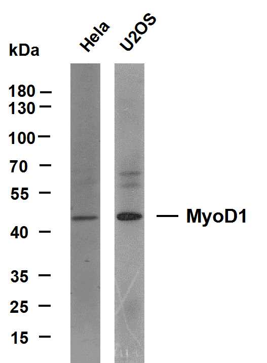
- Various whole cell lysates were separated by 10% SDS-PAGE, and the membrane was blotted with anti-MyoD1(ABT-MYOD1) antibody. The HRP-conjugated Goat anti-Mouse IgG(H + L) antibody was used to detect the antibody. Lane 1: Hela Lane 2: U2OS
.jpg)
- Immunohistochemical analysis of paraffin-embedded Rhabdomyosarcoma. 1, Antibody was diluted at 1:200(4° overnight). 2, Citrate buffer of pH6.0 was used for antigen retrieval. 3,Secondary antibody was diluted at 1:200(room temperature, 30min).
_wb.jpg)
- Western blot analysis of MyoD1Antibody at 1:1000 dilution.


