Keratin-pan Polyclonal Antibody
- Catalog No.:YT5870
- Applications:WB;ELISA;IHC
- Reactivity:Human;Mouse;Rat
- Target:
- Cytokeratin-pan
- Gene Name:
- Keratin
- Protein Name:
- Keratin
- Human Gene Id:
- 3849
- Human Swiss Prot No:
- P35908/Q01546/P12035/P13647/P02538/P04259/P48668/Q3SY84/Q14CN4/Q86Y46/Q7RTS7/O95678/Q5XKE5/P08729/P05787/Q9NSB2
- Immunogen:
- Synthetic peptide from human protein at AA range: 150-220
- Specificity:
- The antibody detects endogenous Keratin-pan
- Formulation:
- Liquid in PBS containing 50% glycerol, 0.5% BSA and 0.02% sodium azide.
- Source:
- Polyclonal, Rabbit,IgG
- Dilution:
- WB 1:500-2000;IHC 1:50-300; ELISA 2000-20000
- Purification:
- The antibody was affinity-purified from rabbit antiserum by affinity-chromatography using epitope-specific immunogen.
- Concentration:
- 1 mg/ml
- Storage Stability:
- -15°C to -25°C/1 year(Do not lower than -25°C)
- Observed Band(KD):
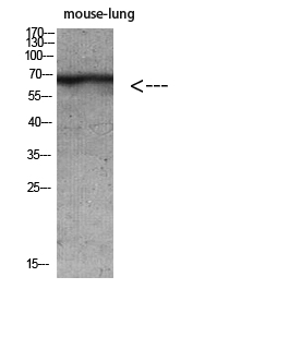
- 70kD
- Background:
- keratin 2(KRT2) Homo sapiens The protein encoded by this gene is a member of the keratin gene family. The type II cytokeratins consist of basic or neutral proteins which are arranged in pairs of heterotypic keratin chains coexpressed during differentiation of simple and stratified epithelial tissues. This type II cytokeratin is expressed largely in the upper spinous layer of epidermal keratinocytes and mutations in this gene have been associated with bullous congenital ichthyosiform erythroderma. The type II cytokeratins are clustered in a region of chromosome 12q12-q13. [provided by RefSeq, Jul 2008],
- Function:
- developmental stage:Synthesized during maturation of epidermal keratinocytes and localized in the upper intermediate cells of fetal skin. Earliest expression is at 10 weeks in the developing embryo in the presumptive nail bed of developing digits, shifting to the proximal nail fold by 13.5 weeks. At 12.5 weeks, detected in scattered cells of the intermediate layer of trunk skin. At 19.3 weeks, regional expression patterns were observed in upper intermediate keratinocytes of cheek, trunk, dorsal and ventral knee, elbow and dorsal hand. Distal areas around the periumbilical region showed increased number of positive cells and by 15 weeks is expressed in small groups of cells in the fetal hair follicles.,disease:Defects in KRT2 are a cause of ichthyosis bullosa of Siemens (IBS) [MIM:146800]. IBS is a rare autosomal dominant skin disorder displaying a type of epidermolytic hyperkeratosis cha
- Subcellular Location:
- extracellular space,nucleus,cytoplasm,Golgi apparatus,intermediate filament,membrane,keratin filament,intermediate filament cytoskeleton,extracellular exosome,
- Expression:
- Expressed in the upper spinous and granular suprabasal layers of normal adult epidermal tissues from most body sites including thigh, breast nipple, foot sole, penile shaft and axilla. Not present in foreskin, squamous metaplasias and carcinomas. Expression in hypertrophic and keloid scars begins in the deepest suprabasal layer. Weakly expressed in normal gingiva and tongue, however expression is induced in benign keratoses of lingual mucosa and in mild-to-moderate oral dysplasia with orthokeratinization.
- June 19-2018
- WESTERN IMMUNOBLOTTING PROTOCOL
- June 19-2018
- IMMUNOHISTOCHEMISTRY-PARAFFIN PROTOCOL
- June 19-2018
- IMMUNOFLUORESCENCE PROTOCOL
- September 08-2020
- FLOW-CYTOMEYRT-PROTOCOL
- May 20-2022
- Cell-Based ELISA│解您多样本WB检测之困扰
- July 13-2018
- CELL-BASED-ELISA-PROTOCOL-FOR-ACETYL-PROTEIN
- July 13-2018
- CELL-BASED-ELISA-PROTOCOL-FOR-PHOSPHO-PROTEIN
- July 13-2018
- Antibody-FAQs
- Products Images

- Western blot analysis of mouse-lung mouse-brain mouse-kidney mouse-heart lysate, antibody was diluted at 500. Secondary antibody(catalog#:RS0002) was diluted at 1:20000

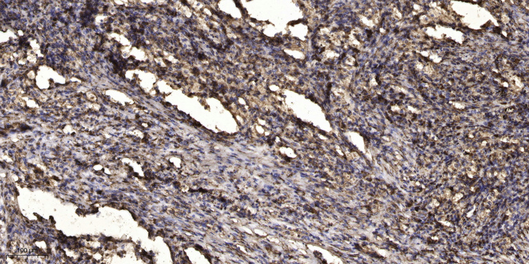
- Immunohistochemical analysis of paraffin-embedded human Squamous cell carcinoma of lung. 1, Antibody was diluted at 1:200(4° overnight). 2, Tris-EDTA,pH9.0 was used for antigen retrieval. 3,Secondary antibody was diluted at 1:200(room temperature, 45min).


