Glut4 Polyclonal Antibody
- Catalog No.:YT5523
- Applications:WB;IHC;IF;ELISA
- Reactivity:Human;Mouse;Rat
- Target:
- Glut4
- Fields:
- >>FoxO signaling pathway;>>AMPK signaling pathway;>>Insulin signaling pathway;>>Adipocytokine signaling pathway;>>Type II diabetes mellitus;>>Insulin resistance;>>Diabetic cardiomyopathy
- Gene Name:
- SLC2A4
- Protein Name:
- Solute carrier family 2 facilitated glucose transporter member 4
- Human Gene Id:
- 6517
- Human Swiss Prot No:
- P14672
- Mouse Gene Id:
- 20528
- Mouse Swiss Prot No:
- P14142
- Rat Gene Id:
- 25139
- Rat Swiss Prot No:
- P19357
- Immunogen:
- The antiserum was produced against synthesized peptide derived from the N-terminal region of human SLC2A4. AA range:21-70
- Specificity:
- Glut4 Polyclonal Antibody detects endogenous levels of Glut4 protein.
- Formulation:
- Liquid in PBS containing 50% glycerol, 0.5% BSA and 0.02% sodium azide.
- Source:
- Polyclonal, Rabbit,IgG
- Dilution:
- WB 1:500 - 1:2000. IHC: 1:100-300 ELISA: 1:20000. IF 1:100-300 Not yet tested in other applications.
- Purification:
- The antibody was affinity-purified from rabbit antiserum by affinity-chromatography using epitope-specific immunogen.
- Concentration:
- 1 mg/ml
- Storage Stability:
- -15°C to -25°C/1 year(Do not lower than -25°C)
- Other Name:
- SLC2A4;GLUT4;Solute carrier family 2, facilitated glucose transporter member 4;Glucose transporter type 4, insulin-responsive;GLUT-4
- Observed Band(KD):
- 56kD
- Background:
- This gene is a member of the solute carrier family 2 (facilitated glucose transporter) family and encodes a protein that functions as an insulin-regulated facilitative glucose transporter. In the absence of insulin, this integral membrane protein is sequestered within the cells of muscle and adipose tissue. Within minutes of insulin stimulation, the protein moves to the cell surface and begins to transport glucose across the cell membrane. Mutations in this gene have been associated with noninsulin-dependent diabetes mellitus (NIDDM). [provided by RefSeq, Jul 2008],
- Function:
- disease:Defects in SLC2A4 may be a cause of noninsulin-dependent diabetes mellitus (NIDDM) [MIM:125853]. Defects in SLC2A4 may be a cause of certain post-receptor defects in NIDDM. The variant in position Ile-383 is found in a small number of NIDDM patients, but seems not to be found in nondiabetic subjects.,function:Insulin-regulated facilitative glucose transporter.,miscellaneous:Insulin-stimulated phosphorylation of TBC1D4 is required for GLUT4 translocation.,online information:GLUT4 entry,PTM:Sumoylated.,similarity:Belongs to the major facilitator superfamily. Sugar transporter (TC 2.A.1.1) family. Glucose transporter subfamily.,subcellular location:Localizes primarily to the perinuclear region, undergoing continued recycling to the plasma membrane where it is rapidly reinternalized. The dileucine internalization motif is critical for intracellular sequestration.,subunit:Binds to DAX
- Subcellular Location:
- Cell membrane ; Multi-pass membrane protein . Endomembrane system ; Multi-pass membrane protein . Cytoplasm, perinuclear region . Localizes primarily to the perinuclear region, undergoing continued recycling to the plasma membrane where it is rapidly reinternalized (PubMed:8300557). The dileucine internalization motif is critical for intracellular sequestration (PubMed:8300557). Insulin stimulation induces translocation to the cell membrane (By similarity). .
- Expression:
- Skeletal and cardiac muscles; brown and white fat.
Structure characterization and anti-fatigue activity of an acidic polysaccharide from Panax ginseng C. A. Meyer
Melatonin treatment improves human umbilical cord mesenchymal stem cell therapy in a mouse model of type II diabetes mellitus via the PI3K/AKT signaling pathway. Stem Cell Research & Therapy2022 Dec;13(1):1-15. Mouse 1: 200 liver, pancreas, white adipose tissue
Melatonin treatment improves human umbilical cord mesenchymal stem cell therapy in a mouse model of type II diabetes mellitus via the PI3K/AKT signaling pathway. Stem Cell Research & Therapy2022 Dec;13(1):1-15. Human,Mouse 1:1200 liver tissue hUC-MSC
Targeting glycometabolic reprogramming to restore the sensitivity of leukemia drug-resistant K562/ADM cells to adriamycin. LIFE SCIENCES Life Sci. 2018 Dec;215:1 WB Human 1:1000 K562 cell
Bawei Chenxiang Wan Ameliorates Cardiac Hypertrophy by Activating AMPK/PPAR-α Signaling Pathway Improving Energy Metabolism. Frontiers in Pharmacology Front Pharmacol. 2021 Jun;0:1314 WB Rat Myocardial tissue
CD38 affects the biological behavior and energy metabolism of nasopharyngeal carcinoma cells. INTERNATIONAL JOURNAL OF ONCOLOGY Int J Oncol. 2019 Feb;54(2):585-599 WB Human 1:1000 5-8F cell
PM2.5 aggravates diabetes via the systemically activated IL-6-mediated STAT3/SOCS3 pathway in rats’ liver. ENVIRONMENTAL POLLUTION Environ Pollut. 2020 Jan;256:113342 WB Rat skeletal muscles,abdominal fat, liver
Preventative effects of resveratrol and estradiol on streptozotocin-induced diabetes in ovariectomized mice and the related mechanisms. PLoS One 2018 Oct 01 WB,IHC Mouse Skeletal muscle,myocardial tissues
Glycometabolic adaptation mediates the insensitivity of drug-resistant K562/ADM leukaemia cells to adriamycin via the AKT-mTOR/c-Myc signalling pathway. Molecular Medicine Reports Mol Med Rep. 2017 Apr;15(4):1869-1876 WB Human 1:1000 K562/ADM cell
Long, Min-hui, et al. "PM2. 5 aggravates diabetes via the systemically activated IL-6-mediated STAT3/SOCS3 pathway in rats’ liver." Environmental Pollution 256 (2020): 113342.
Bawei Chenxiang Wan Ameliorates Cardiac Hypertrophy by Activating AMPK/PPAR-α Signaling Pathway Improving Energy Metabolism. Frontiers in Pharmacology Front Pharmacol. 2021 Jun;0:1314 WB Rat Myocardial tissue
Acidic polysaccharides of Mountain Cultivated Ginseng: The potential source of anti-fatigue nutrients Journal of Functional Foods Yang Yu WB Mouse
The role of chemerin in the regulation of cGAS-STING pathway in gestational diabetes mellitus placenta FASEB JOURNAL Ling Feng IF Human HTR-8/SVneo cell
Soy protein compared with whey protein ameliorates insulin resistance by regulating lipid metabolism, AMPK/mTOR pathway and gut microbiota in high-fat diet-fed mice. Duo Li WB Mouse liver,gastrocnemius muscle
- June 19-2018
- WESTERN IMMUNOBLOTTING PROTOCOL
- June 19-2018
- IMMUNOHISTOCHEMISTRY-PARAFFIN PROTOCOL
- June 19-2018
- IMMUNOFLUORESCENCE PROTOCOL
- September 08-2020
- FLOW-CYTOMEYRT-PROTOCOL
- May 20-2022
- Cell-Based ELISA│解您多样本WB检测之困扰
- July 13-2018
- CELL-BASED-ELISA-PROTOCOL-FOR-ACETYL-PROTEIN
- July 13-2018
- CELL-BASED-ELISA-PROTOCOL-FOR-PHOSPHO-PROTEIN
- July 13-2018
- Antibody-FAQs
- Products Images

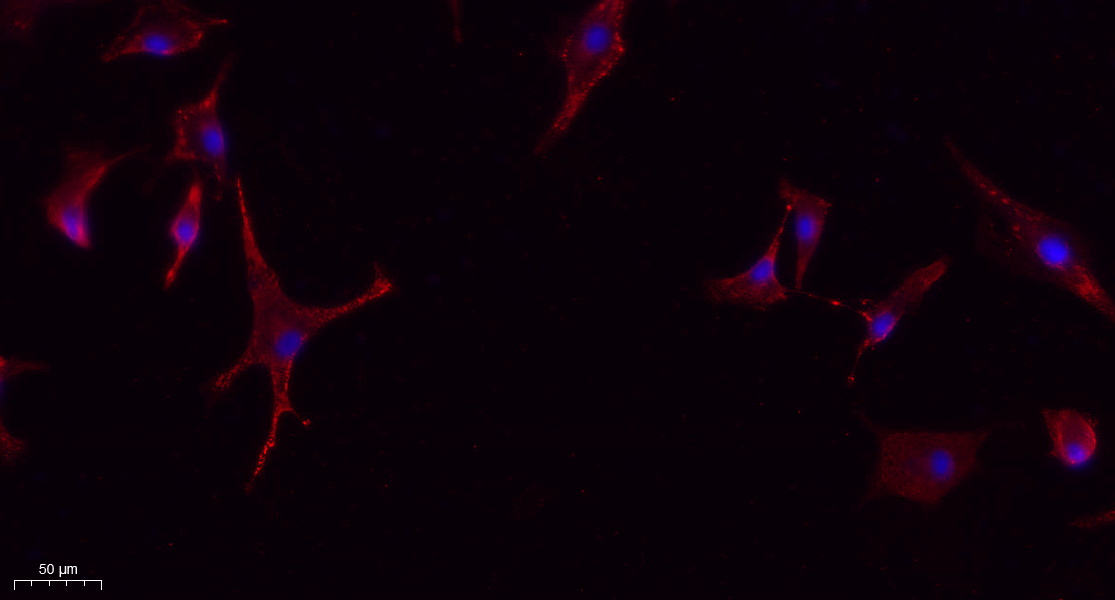
- Immunofluorescence analysis of A549. 1,primary Antibody(red) was diluted at 1:200(4°C overnight). 2, Goat Anti Rabbit IgG (H&L) - Alexa Fluor 594 Secondary antibody was diluted at 1:1000(room temperature, 50min).3, Picture B: DAPI(blue) 10min.
.jpg)
- Long, Min-hui, et al. "PM2. 5 aggravates diabetes via the systemically activated IL-6-mediated STAT3/SOCS3 pathway in rats’ liver." Environmental Pollution 256 (2020): 113342.

- Immunohistochemical analysis of paraffin-embedded Rat-lung tissue. 1,Glut4 Polyclonal Antibody was diluted at 1:200(4°C,overnight). 2, Sodium citrate pH 6.0 was used for antibody retrieval(>98°C,20min). 3,Secondary antibody was diluted at 1:200(room tempeRature, 30min). Negative control was used by secondary antibody only.

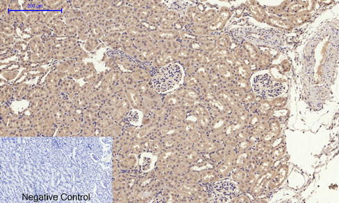
- Immunohistochemical analysis of paraffin-embedded Rat-kidney tissue. 1,Glut4 Polyclonal Antibody was diluted at 1:200(4°C,overnight). 2, Sodium citrate pH 6.0 was used for antibody retrieval(>98°C,20min). 3,Secondary antibody was diluted at 1:200(room tempeRature, 30min). Negative control was used by secondary antibody only.

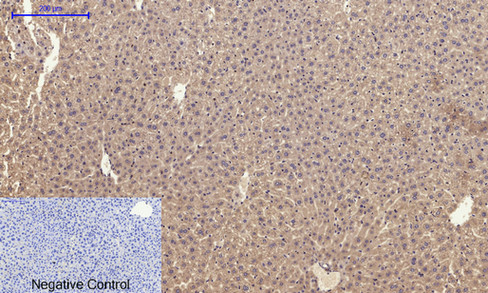
- Immunohistochemical analysis of paraffin-embedded Mouse-liver tissue. 1,Glut4 Polyclonal Antibody was diluted at 1:200(4°C,overnight). 2, Sodium citrate pH 6.0 was used for antibody retrieval(>98°C,20min). 3,Secondary antibody was diluted at 1:200(room tempeRature, 30min). Negative control was used by secondary antibody only.

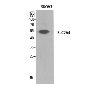
- Western Blot analysis of SKOV3 cells using Glut4 Polyclonal Antibody. Secondary antibody(catalog#:RS0002) was diluted at 1:20000


