Adducin α/β (phospho Ser726/713) Polyclonal Antibody
- Catalog No.:YP0854
- Applications:WB;IHC;IF;ELISA
- Reactivity:Human;Mouse;Rat
- Target:
- Adducin α/β
- Gene Name:
- ADD1/ADD2
- Protein Name:
- Alpha-adducin/Beta-adducin
- Human Gene Id:
- 118/119
- Human Swiss Prot No:
- P35611/P35612
- Mouse Gene Id:
- 11518/11519
- Rat Gene Id:
- 24170/24171
- Rat Swiss Prot No:
- Q63028/Q05764
- Immunogen:
- The antiserum was produced against synthesized peptide derived from human ADD1 around the phosphorylation site of Ser726. AA range:688-737
- Specificity:
- Phospho-Adducin α/β (S726/713) Polyclonal Antibody detects endogenous levels of Adducin α/β protein only when phosphorylated at S726/713.
- Formulation:
- Liquid in PBS containing 50% glycerol, 0.5% BSA and 0.02% sodium azide.
- Source:
- Polyclonal, Rabbit,IgG
- Dilution:
- WB 1:500 - 1:2000. IHC 1:100 - 1:300. IF 1:200 - 1:1000. ELISA: 1:40000. Not yet tested in other applications.
- Purification:
- The antibody was affinity-purified from rabbit antiserum by affinity-chromatography using epitope-specific immunogen.
- Concentration:
- 1 mg/ml
- Storage Stability:
- -15°C to -25°C/1 year(Do not lower than -25°C)
- Other Name:
- ADD1;ADDA;Alpha-adducin;Erythrocyte adducin subunit alpha;ADD2;ADDB;Beta-adducin;Erythrocyte adducin subunit beta
- Observed Band(KD):
- 80kD
- Background:
- adducin 1(ADD1) Homo sapiens Adducins are a family of cytoskeleton proteins encoded by three genes (alpha, beta, gamma). Adducin is a heterodimeric protein that consists of related subunits, which are produced from distinct genes but share a similar structure. Alpha- and beta-adducin include a protease-resistant N-terminal region and a protease-sensitive, hydrophilic C-terminal region. Alpha- and gamma-adducins are ubiquitously expressed. In contrast, beta-adducin is expressed at high levels in brain and hematopoietic tissues. Adducin binds with high affinity to Ca(2+)/calmodulin and is a substrate for protein kinases A and C. Alternative splicing results in multiple variants encoding distinct isoforms; however, not all variants have been fully described. [provided by RefSeq, Jul 2008],
- Function:
- alternative products:Additional isoforms seem to exist,domain:Each subunit is comprised of three regions: a NH2-terminal protease-resistant globular head region, a short connecting subdomain, and a protease-sensitive tail region.,function:Membrane-cytoskeleton-associated protein that promotes the assembly of the spectrin-actin network. Binds to calmodulin.,PTM:The N-terminus is blocked.,similarity:Belongs to the aldolase class II family. Adducin subfamily.,subunit:Heterodimer of an alpha and a beta subunit or an alpha and a gamma subunit. Binds ROCK1.,tissue specificity:Expressed in all tissues. Found in much higher levels in reticulocytes than the beta subunit.,
- Subcellular Location:
- Cytoplasm, cytoskeleton. Cell membrane; Peripheral membrane protein; Cytoplasmic side.
- Expression:
- Expressed in all tissues. Found in much higher levels in reticulocytes than the beta subunit.
- June 19-2018
- WESTERN IMMUNOBLOTTING PROTOCOL
- June 19-2018
- IMMUNOHISTOCHEMISTRY-PARAFFIN PROTOCOL
- June 19-2018
- IMMUNOFLUORESCENCE PROTOCOL
- September 08-2020
- FLOW-CYTOMEYRT-PROTOCOL
- May 20-2022
- Cell-Based ELISA│解您多样本WB检测之困扰
- July 13-2018
- CELL-BASED-ELISA-PROTOCOL-FOR-ACETYL-PROTEIN
- July 13-2018
- CELL-BASED-ELISA-PROTOCOL-FOR-PHOSPHO-PROTEIN
- July 13-2018
- Antibody-FAQs
- Products Images
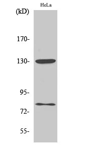
- Western Blot analysis of various cells using Phospho-Adducin α/β (S726/713) Polyclonal Antibody diluted at 1:1000
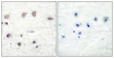
- Immunohistochemical analysis of paraffin-embedded Human breast cancer. Antibody was diluted at 1:100(4° overnight). High-pressure and temperature Tris-EDTA,pH8.0 was used for antigen retrieval. Negetive contrl (right) obtaned from antibody was pre-absorbed by immunogen peptide.
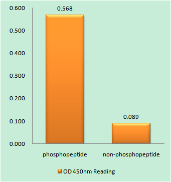
- Enzyme-Linked Immunosorbent Assay (Phospho-ELISA) for Immunogen Phosphopeptide (Phospho-left) and Non-Phosphopeptide (Phospho-right), using ADD1 (Phospho-Ser726) Antibody
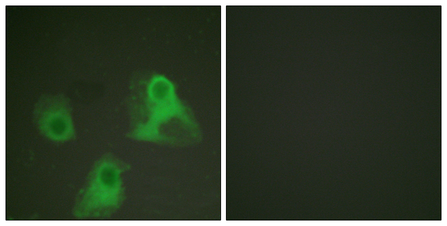
- Immunofluorescence analysis of HeLa cells, using ADD1 (Phospho-Ser726) Antibody. The picture on the right is blocked with the phospho peptide.
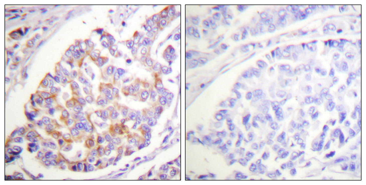
- Immunohistochemistry analysis of paraffin-embedded human breast carcinoma, using ADD1 (Phospho-Ser726) Antibody. The picture on the right is blocked with the phospho peptide.
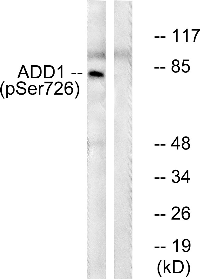
- Western blot analysis of lysates from HeLa cells treated with Forskolin 40nM 30', using ADD1 (Phospho-Ser726) Antibody. The lane on the right is blocked with the phospho peptide.


