Rb (phospho Thr826) Polyclonal Antibody
- Catalog No.:YP0556
- Applications:WB;IHC;IF;ELISA
- Reactivity:Human;Mouse;Rat
- Target:
- Rb
- Fields:
- >>Endocrine resistance;>>Cell cycle;>>Cellular senescence;>>Cushing syndrome;>>Hepatitis C;>>Hepatitis B;>>Human cytomegalovirus infection;>>Human papillomavirus infection;>>Human T-cell leukemia virus 1 infection;>>Kaposi sarcoma-associated herpesvirus infection;>>Epstein-Barr virus infection;>>Pathways in cancer;>>Viral carcinogenesis;>>Chemical carcinogenesis - receptor activation;>>Pancreatic cancer;>>Glioma;>>Prostate cancer;>>Melanoma;>>Bladder cancer;>>Chronic myeloid leukemia;>>Small cell lung cancer;>>Non-small cell lung cancer;>>Breast cancer;>>Hepatocellular carcinoma;>>Gastric cancer
- Gene Name:
- RB1
- Protein Name:
- Retinoblastoma-associated protein
- Human Gene Id:
- 5925
- Human Swiss Prot No:
- P06400
- Mouse Gene Id:
- 19645
- Mouse Swiss Prot No:
- P13405
- Rat Gene Id:
- 24708
- Rat Swiss Prot No:
- P33568
- Immunogen:
- The antiserum was produced against synthesized peptide derived from human Retinoblastoma around the phosphorylation site of Thr826. AA range:601-650
- Specificity:
- Phospho-Rb (T826) Polyclonal Antibody detects endogenous levels of Rb protein only when phosphorylated at T826.
- Formulation:
- Liquid in PBS containing 50% glycerol, 0.5% BSA and 0.02% sodium azide.
- Source:
- Polyclonal, Rabbit,IgG
- Dilution:
- WB 1:500 - 1:2000. IHC 1:100 - 1:300. IF 1:200 - 1:1000. ELISA: 1:10000. Not yet tested in other applications.
- Purification:
- The antibody was affinity-purified from rabbit antiserum by affinity-chromatography using epitope-specific immunogen.
- Concentration:
- 1 mg/ml
- Storage Stability:
- -15°C to -25°C/1 year(Do not lower than -25°C)
- Other Name:
- RB1;Retinoblastoma-associated protein;p105-Rb;pRb;Rb;pp110
- Observed Band(KD):
- 110kD
- Background:
- The protein encoded by this gene is a negative regulator of the cell cycle and was the first tumor suppressor gene found. The encoded protein also stabilizes constitutive heterochromatin to maintain the overall chromatin structure. The active, hypophosphorylated form of the protein binds transcription factor E2F1. Defects in this gene are a cause of childhood cancer retinoblastoma (RB), bladder cancer, and osteogenic sarcoma. [provided by RefSeq, Jul 2008],
- Function:
- disease:Defects in RB1 are a cause of bladder cancer [MIM:109800].,disease:Defects in RB1 are a cause of osteogenic sarcoma [MIM:259500].,disease:Defects in RB1 are the cause of childhood cancer retinoblastoma (RB) [MIM:180200]. RB is a congenital malignant tumor that arises from the nuclear layers of the retina. It occurs in about 1:20'000 live births and represents about 2% of childhood malignancies. It is bilateral in about 30% of cases. Although most RB appear sporadically, about 20% are transmitted as an autosomal dominant trait with incomplete penetrance. The diagnosis is usually made before the age of 2 years when strabismus or a gray to yellow reflex from pupil ("cat eye") is investigated.,function:Key regulator of entry into cell division that acts as a tumor suppressor. Acts as a transcription repressor of E2F1 target genes. The underphosphorylated, active form of RB1 interacts
- Subcellular Location:
- Nucleus . During keratinocyte differentiation, acetylation by KAT2B/PCAF is required for nuclear localization. .
- Expression:
- Expressed in the retina. Expressed in foreskin keratinocytes (at protein level) (PubMed:20940255).
- June 19-2018
- WESTERN IMMUNOBLOTTING PROTOCOL
- June 19-2018
- IMMUNOHISTOCHEMISTRY-PARAFFIN PROTOCOL
- June 19-2018
- IMMUNOFLUORESCENCE PROTOCOL
- September 08-2020
- FLOW-CYTOMEYRT-PROTOCOL
- May 20-2022
- Cell-Based ELISA│解您多样本WB检测之困扰
- July 13-2018
- CELL-BASED-ELISA-PROTOCOL-FOR-ACETYL-PROTEIN
- July 13-2018
- CELL-BASED-ELISA-PROTOCOL-FOR-PHOSPHO-PROTEIN
- July 13-2018
- Antibody-FAQs
- Products Images

- Western blot analysis of RAT-musle using p-Rb (T826) antibody. Antibody was diluted at 1:500 cells nucleus extracted by Minute TM Cytoplasmic and Nuclear Fractionation kit (SC-003,Inventbiotech,MN,USA).

- Immunohistochemical analysis of paraffin-embedded Human brain. Antibody was diluted at 1:100(4° overnight). High-pressure and temperature Tris-EDTA,pH8.0 was used for antigen retrieval. Negetive contrl (right) obtaned from antibody was pre-absorbed by immunogen peptide.

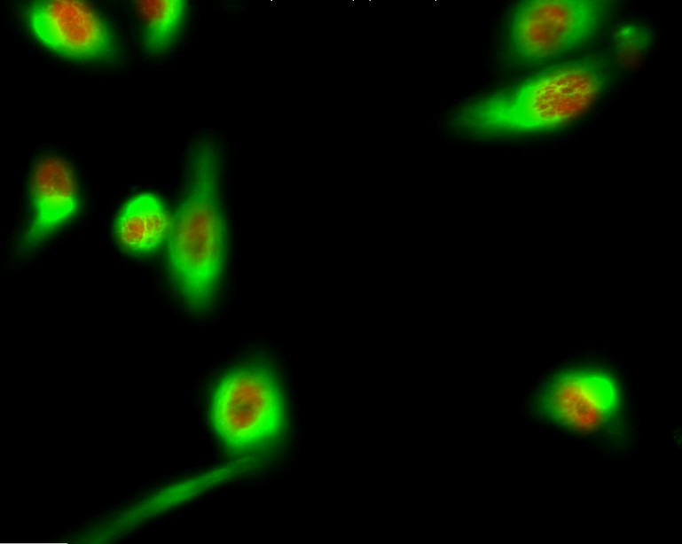
- Immunofluorescence analysis of COS7 cells, using Retinoblastoma (Phospho-Thr826) Antibody. The picture on the right is blocked with the phospho peptide.

- Western blot analysis of lysates from HepG2 cells treated with nocodazole 1ug/ml 16h, using Retinoblastoma (Phospho-Thr826) Antibody. The lane on the right is blocked with the phospho peptide.

